Orthopedic Surgery Enters the Modern Age on a Chance Observation
Roy A. Meals, MD
For thousands of years, bone setters and doctors could not accurately diagnosis broken bones or differentiate such injuries from joint dislocations and torn ligaments. That changed with a chance discovery 125 years ago this month. Subsequently, doctors began using the new discovery to discount their long-held assumptions and were able to accurately diagnose skeletal diseases.
In his darkened laboratory on November 8, 1895, a German mechanical engineer and physicist, Wilhelm Röntgen, electrified a vacuum tube and happened to observe a strange glow coming from a nearby card coated with a photosensitive chemical. He placed his hand between the vacuum tube and the card and an image of his hand appeared. Over the following weeks he ate and slept in his laboratory and studied this unknown ray, which he labeled “X,” the mathematical symbol for an unknown. 
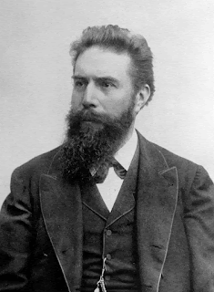
Wilhelm Röntgen (Wikipedia)
Röntgen discovered that X-rays passed through books, no matter how thick, and that coins cast a shadow on the photosensitive board. Röntgen shared the secret with his wife, who allowed him to take a fifteen-minute exposure of her hand, the first orthopedic X-ray. When she saw the image of her hand skeleton, she exclaimed, “I have seen my death.” Far more broadly, she was witnessing the advent of diagnostic radiology and modern orthopedic surgery.
A week later, Röntgen presented his findings in a paper titled, “On a New Type of Rays.” This caught the immediate attention of physicists, who alerted the lay press. The discovery made the front-page headline news within a week of Röntgen’s public presentation.
At the time, vacuum tubes were well known and easy to make. After Röntgen’s discovery and announcement, many investigators contributed to the understanding and practical applications of X-rays. Interest was intense and advances were rapid. Less than three months after Röntgen’s public announcement, an enterprising electrical contractor and avid photographer opened a laboratory that offered diagnostic services. 
X-ray of Bertha's hand (Wikipedia)
Röntgen received the Nobel Prize in 1901, the first one ever awarded for physics. Röntgen not only gave the reward money to his university, he also refused to take out patents on his discovery to allow for wide-spread application.
I suppose when X-rays were in their infancy, patients were asking, “Now that you have finished obtaining a thorough medical history, performing a careful physical examination, and telling me that you know with assurance what is wrong, aren’t you going to order an X-ray, Doctor?” This question implied a lack of trust in the doctor’s diagnosis unless he threw in a high-tech, oh-so-modern X-ray evaluation. Gradually, doctors and patients came to understand when an X-ray study could help make the diagnosis or plan treatment and when one would be superfluous. For instance, today it is intuitive that a sore tooth most likely deserves an X-ray while a sore throat does not. In general, X-rays reveal calcium-rich structures—those containing enough calcium to cast a shadow in the X-ray beam. Examples are bones, teeth, hardened arteries, and kidney stones.
Doctors have learned to order X-rays with some caution because radiation damages living tissues and their DNA. That fact required discovery, and the harmful effects of early X-ray examinations were slow to reveal themselves. Since X-rays could not be seen or felt, investigators had no reason to consider them harmful. Both Nicola Tesla and Thomas Edison experimented with X-rays, and both observed that their eyes became irritated; but neither drew a connection between the radiation and their symptoms.
For convenience, dentists originally held the film inside the patient’s mouth with their fingers when shooting dental X-rays. Decades later the skin on their hands dried, cracked, and became cancerous. Nowadays the radiology tech steps behind a lead shield before shooting the film, and there are generally accepted standards for how much radiation a person can receive on an annual and lifetime basis without incurring undue risk.
Despite our best efforts, we cannot avoid radiation exposure entirely. Some comes naturally from the sun and some from the ground. We get more during a plane flight because the thinner air at high altitude blocks less of the sun’s radiation. This fact poses a major, unsolved problem for interplanetary travel because of the absence of Earth’s radiation-shielding atmosphere and because of the impracticality of armoring spaceships with lead.
Most would agree, however, that the potential benefits of a timely chest X-ray or mammogram far outweigh the risks. Even an occasional and judiciously planned CT scan may help maintain or restore one’s health; but we should avoid advice such as, “I don’t have a clue about what’s wrong, so let’s get a CT scan.” A second opinion is safer. Remember, it took decades for the damaged DNA in dentists to turn into skin cancers. Similarly, we should avoid being our own doctor and proclaiming, “I would just feel better, Doctor, if you ordered a CT scan.”
Röntgen discovered his new type of ray a few decades after the introduction of general anesthesia and the acceptance of aseptic surgical techniques. These developments, along with the invention of stainless steel, ushered into the modern era orthopedic surgery and the practicality of operative fixation of fractures. Looking ahead 125 years, fractures will still exist. Arthritis and osteoporosis may be fully preventable. Bone imaging techniques will be even more sophisticated than those in current use. X-ray imaging may be obsolete, replaced completely by magnetic resonance imaging, ultrasound, or some yet-to-be-discovered alternative. Nevertheless, Röntgen’s discovery and its enduring 125-year legacy deserves recognition and respect.
Bradley, William. “History of Medical Imaging.” Proceedings of the American Philosophical Society 152, no. 3 (2008): 349–61.
“Radiation Risk from Medical Imaging.” Accessed November 6, 2020. https://www.health.harvard.edu/cancer/radiation-risk-from-medical-imaging
Röntgen, William. “Ueber eine neue Art von Strahlen. (On a New Kind of Rays.)” In Classics of Orthopaedics. Edited by Edgar Bick, 278–84. Philadelphia: Lippincott, 1976.
Dr. Meals blogs at www.aboutbone.com and has recently published Bones, Inside and Out (WW Norton, 2020)

No comments:
Post a Comment
Comments are welcome. Especially new knowledge, insights, and sources of further information.