Wednesday, December 13, 2017

CHRISTMAS, DICKENS, AND MEDICINE

     “God bless us, every one”, the final words spoken by Tiny Tim in Charles Dickens’ A Christmas Carol, still echo after over 150 years. It’s a tale of the power of Christmas to soften up a “squeezing, wrenching, grasping, scraping, clutching, covetous old sinner” like Ebeneezer Scrooge.
     Tiny Tim, Bob Cratchit’s little son, has aroused medical curiosity. He is depicted as small for his age and carried on his father’s shoulder. He “bore a little crutch and had his limbs supported by an iron frame”, and had a “withered little hand”. He often sits by himself and “thinks the strangest things you ever heard”, though not irrational thoughts. 
Tiny Tim on Bob Cratchit's shoulder (Wikipedia)
     What was wrong with Tiny Tim? The story does not say but there was fluctuation in his weakness and eventually the boy recovered. Donald W. Lewis, a pediatric neurologist, after ruling out tuberculosis of the spine and rickets by events in the story, made a case for renal tubular acidosis, favoring type I RTA. This disorder, by producing increased body acidification leads to growth retardation, osteomalacia, bone pain and pathologic fractures. A review of British pediatric texts of Dickens' time revealed that general treatments for almost any illness included fresh air and sunshine, a balanced diet, fish liver oils, and tonics for digestion. In Tim’s case treatment for rickets or TB might have been added, and rickets was managed the same way as scrofula. Such patients were believed to have an excess of acid and received alkaline carbonates such as bicarbonate of soda or other carbonates. This combination, especially the alkalinizing effect of bicarbonates, Dr. Lewis believed, could have led to Tiny Tim’s recovery.
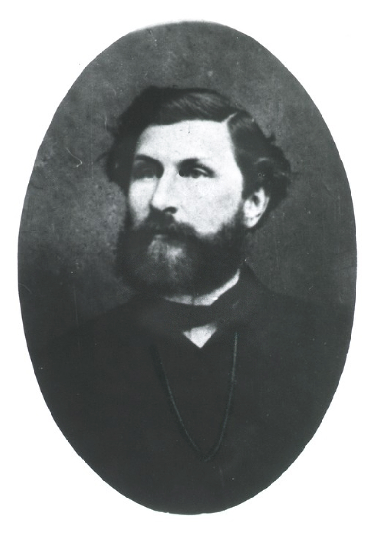
     Medical problems pop up in much of Dickens’ fiction. (Someone even wrote a book about it.) Just to mention a few, we read about the fat, lethargic boy Joe in the Pickwick Papers, believed to have a “Pickwickian syndrome”. Other stories mention ataxic gait, gout, erysipelas, typhoid, dwarfism, opium use, and additional problems.
Charles Dickens, Photo of George Herbert Watkins
(Wikipedia)
     Children populated many of his novels and he took great interest in their welfare. He castigated child labor conditions at a time when children as young as seven worked in mines and other dangerous jobs. In 1850 in London about one half of all deaths were in children and yet there was no children’s hospital. Through the efforts of Dr. Charles West, assisted by Dr. Henry Bence-Jones (of the myeloma protein) and others, the Hospital for Sick Children went up in 1852 on Great Ormond Street, London, in a mansion that previously housed Queen Anne’s physician, Dr. Richard Mead, and his 100,000-volume library. Dickens raised funds for it by public speaking and a reading of A Christmas Carol.  
     The London of Dickens was pretty filthy. Thick smog all but obliterated sunlight much of the time. The gargantuan clouds of smoke pouring out from soft coal fires joined with Thames Valley mist to darken the streets, irritate the eyes, and create havoc for asthmatics. In poorer areas sanitation was almost absent and the Thames itself was a depository of tons of sewage. It took the “great stink” of 1858 to force members of Parliament, based on the Thames and literally holding handkerchiefs over their noses, to pass a bill authorizing a citywide sewage project. Housing was cramped, food often scarce, and water impure. People, including children, often walked miles to work.
     Dickens took an interest in public health. He was an anti-contagionist, attributing diseases of poverty to miasmas arising 
Joseph Southwood-Smith (Wellcome Library)
from unsanitary conditions. He befriended sanitarians such as Dr. Joseph Southwood-Smith, the famous public health advocate, fever expert, and co-believer in miasmas, and Edwin Chadwick, the
lawyer who authored the Report on the Sanitary Condition of the Labouring Population in Great Britain. The Report was a bombshell exposé that helped usher in better sanitation. Dickens supported their work with his own writings.
     Dickens was also close to Thomas Wakley. Wakley was a
Thomas Wakley (Wikipedia)
combative surgeon, reformer, coroner, Member of Parliament, and editor of Lancet at various times in his career. He used the Lancet as a platform for reforming medicine and public health.
     Dickens’ connections to medicine could go on, but that’s enough for now. Good health to all,

                     HAPPY HOLIDAYS
       and     A JOYOUS NEW YEAR.


SOURCES:
Hearn, Michael P., ed. The Annotated  
         Christmas Carol, by Charles Dickens. 2004.
Tomalin, Claire. Charles Dickens: A Life. 2011.
Flanders, Judith. The Victorian City: Everyday Life in Dickens’
         London. 2012.
Corton, Christine. London Fog: The Biography. 2015.
Cambridge, Nicholas. “From Mr. Pickwick to Tiny Tim: Charles
         Dickens and Medicine”. Lecture at Gresham College. available
Lewis, Donald W. “What was wrong with Tiny Tim?” Am J Dis
         Child 1992; 146(12): 1403-7.
Eysell, Joanne. A Medical Companion to Dickens's Fiction. 2005.


Thursday, November 16, 2017

SKIN DISEASE IN OLD VIENNA

     Imagine a king roaming around his realm in disguise to better know the problems of his subjects. It’s the stuff of a fairy tale, but it actually happened – in Vienna, where the Habsburg emperor Joseph II ruled from 1780-90. A liberal but impulsive monarch inspired by enlightenment ideas, he established in 1784 the Allgemeines Krankenhaus (General Hospital) as part of a larger program to deal with the sick and poor streaming into the city since the start of industrialization.  The hospital that Joseph created, using his own funds, was a makeover of an old almshouse built around several courtyards, redesigned to house about 2000 patients.
Allgemeines Krankenhaus (Wikipedia)
     In the decade 1836-46, under the influence of the enlightened vice-director of the faculty, Ludwig Baron von Türkheim, the medical school attached to the hospital was substantially reformed. Josef Skoda, the great diagnostician, was given his own chest service, and Carl Rokitansky created a pathology department (he eventually performed some 30,000 autopsies). Other specialty divisions were established, making the “second” Vienna Medical School one of the most advanced in the world, drawing on many thousands of patients seen each year at the hospital.
     Patients with skin diseases, before the 1840s, were generally placed in medical departments. Most skin conditions were thought to result from attempts of poisons or “corruptions” from within the body to escape through the surface. Thus skin lesions were often left alone so as not to impede this process. At the Allgemeines Krankenhaus the skin ward was next to Skoda’s chest ward, and Skoda asked one of his brightest graduating students, Ferdinand von Hebra, to take charge of the ward.
     Ferdinand Ritter von Hebra was born in Brünn, Moravia, in 1816. He attended high school at a monastery in Styria, then
(Wikipedia)
medical school in Vienna, graduating in 1841. Josef Skoda arranged for Hebra to be his assistant. Noticing that Hebra took an interest in the neglected dermatology patients Skoda urged him to study the patients, and supplied him with books.
      Hebra absorbed existing literature intensively - in particular the writings of two English physicians, Robert Willan and Thomas Bateman, and the Frenchmen, Jean Louis Alibert and Laurent-Théodore Biett, all of whom had attempted to bring some order out of the confusion of skin lesions. Alibert’s classification took the form of a tree, with the trunk representing the epidermis and dermis, and the branches various groups of diseases. All these classifications fell short, partly due to inadequate knowledge.
Arbre des Dermatoses by Alibert - illustrating
his classification (Wikipedia)
     Over time Hebra concluded that many skin ailments were of local origin and not expressions of poisons escaping from the inner body. This was most evident in the case of scabies, perhaps the most common disease he encountered (he is said to have seen 60,000 cases by 1860). The causative mite, Sarcoptes scabiei, though suspected for many years, had proved difficult to find. It was not until a student of Alibert, Simon Francois Renucci, in 1834 demonstrated the mite consistently (by looking between vesicles rather than inside them) at the St. Louis Hospital, Paris. His demonstration even survived a 300-franc bet that he was wrong.
     Hebra enlarged upon Renucci’s work by infecting himself several times and charting the course and cure of the disease. He did away with bleedings, laxatives, and most medicines taken internally, treatments reflecting the belief of internal disease as a cause. Instead he applied sulfur, in an ointment made from lard and potassium bicarbonate, and noted the necessity of treating all skin but the head. He acknowledged that “quacks and old women” had more sense than doctors in their use of topical therapies.
Vienna Medical Faculty
Hebra(second from left, rear), Skoda (second from left, front),
Rokitansky (center, front) ( from Google Books)
     Hebra’s work on scabies carried great weight in conveying the idea that the skin should be considered a separate organ subject to its own disorders, though he recognized that some skin problems were indeed manifestations of generalized disease. Soothing baths, oils, and sometimes more caustic applications were used, many of them tested systematically, and most internal remedies discarded.
     Hebra advanced the description and organization of dermatologic lesions. Terms such as erythema multiforme, lichen planus, impetigo herpetiformis, and rhinoscleroma are largely due to him. He began lectures on skin disease in his second year and was an exceptionally good lecturer, combining clarity, abundant
Erythema multiform (from Hebra: Atlas
der Hautkrankheiten,
Google Books)
material, and a ready wit. The lectures attracted students from home and abroad. He could also, in Sherlock Holmes fashion, tell a patient's occupation by examining their hands or feet. In 1845 Hebra’s ward was separated from Skoda’s to become an autonomous dermatology service, a moment considered by some as the “birthdate of dermatology” as a specialty. He published many articles and a few books, including a famous atlas of skin diseases. The latter was a collaboration with the artists Anton Elfinger, who was also a successful political cartoonist, and Carl Heitzmann. 

     Heitzmann had studied medicine under
From Atlas der Descriptiven Anatomie des Menschen
by Carl Heitzmann. (Hathi Trust)
both Hebra and Rokitansky and was even being considered as a successor to Rokitansky as chair of pathology. He never had formal art training but nevertheless published a surgical pathology text for medical students and later a two-volume human anatomy text with 600 illustrations that went through 9 editions. After failing to succeed Rokitansky he emigrated to New York where he practiced dermatology, published more papers, and was a founding member of the American Academy of Dermatology.
     An assistant to Hebra was Moritz Kaposi, whose name is
Moritz Kaposi (Wikipedia)
associated with a formerly rare but now common malignancy seen in HIV patients. Kaposi was gifted and hard-working, and finished volume 2 of Hebra’s Handbook of Dermatology after Hebra’s death. He also married the boss’ daughter.
     Ferdinand von Hebra died in 1880 of “dropsy”. A funeral procession a mile and a half long followed his coffin to its final resting place next to Carl Rokitansky. Von Hebra brought dermatology into the modern world, freeing it from many misconceptions and opening it to modern research methods.

SOURCES:
Crissey, J T. and Parish, L C. The Dermatology and Syphilology of the Nineteenth
         Century. 1981, Praeger Scientific. Especially chapter 4.
Lesky, E. The Vienna Medical School of the 19th Century. 1976, Johns Hopkins Univ
        Press.
Finnerud, C W: “Ferdinand von Hebra and the Vienna School of Dermatology”. AMA
        Arch Derm Syph.  1952, 66(2): 223-32.
Everett, M A: “Jean Louis Alibert: The Father of French Dermatology”. Int J Derm
       1984, 23: 351-6.
Neuburger, M. “Die Lehre von den Hautkrankheiten vor Hebra”. Wien Med Wochen 
          1928, 78: 641.
Friedman, R: “The Story of Scabies II”. Medical Life 1934, 41: 426-76. 
Friedman, R: “The Story of Scabies III”. Medical Life 1935, 42: 218-71.
Hackstock, I: “Carl Heitzmann (1836-1896): physician and illustrator”. Int J Derm
         1938, 37: 235-40.
Shelly, W B, and Crissey, J T: Classics in Clinical Dermatology, with Biographical
        Sketches. 1953, Charles Thomas.
Lesky, E. Meilensteine der Wiener Medizin. 1981, Verlag Wilhelm  
        Modrig.


Wednesday, October 18, 2017

A GIANT IN PHYSIOLOGY


     American physiology and medicine owes much to a German pioneer and master teacher of the subject: Carl Ludwig. Ludwig was born in 1816 in the town of Witzenhausen, near Cassel, in the aftermath of the Napoleonic wars. He studied medicine at the University of Marburg, obtaining his degree in 1839. During time as a prosector in the anatomy department there he published a dissertation on the mechanism of urine formation, foreshadowing mechanisms of diffusion and osmosis. A few years later he was full professor of anatomy. 
Carl Ludwig (Wikipedia)
     Ludwig’s ascent occurred during a time when German sciences were hampered by backwardness and a lingering belief in a “vital force” to explain phenomena in living material. Fortunately he was close to the chemist, Robert Bunsen (of the Bunsen burner), who influenced him to think of chemistry and physics as drivers of life processes. Shortly after, in 1847, Ludwig, an inveterate experimenter, invented the kymograph, a revolving drum coated with soot, on which a needle recorded events over time, such as pulse, respiration, etc. Its simplicity and its value in visualizing these processes made it popular in physiology labs worldwide. 
Ludwig's Kymograph (Wikipedia)
     In that same year Ludwig traveled to Berlin where he met Hermann von Helmholtz, Emil du Bois-Reymond, and Ernst von Brücke – all students of Johannes Müller, one of the first physiologists. Müller’s three students and Ludwig formed a quartet that set out to change concepts in physiology and anchor the entire subject in chemistry, physics, and anatomy, rejecting ideas of a “vital” force. They were largely successful.
     Two years later Ludwig went to Zurich as Professor of Anatomy and Physiology. There he devised methods to measure blood flow and blood gases and published the first volume of a new, groundbreaking text, Lehrbuch der Physiologie (textbook of physiology). It was the first “modern” such text and a direct refutation of the “vitalist” theories of biology that postulated that body functions could not be explained by chemistry and physics alone.  
Page from Vol. 1, Lehrbuch der Physiologie
(Hathi Trust)
     After a short time in Vienna (where volume two of his text was published) Ludwig was asked to head a new physiology institute at Leipzig in 1865. This was to be the most modern in the world. It consisted three separate wings, in the shape of an E, for anatomy (and histology), chemistry, and (biological) physics, with a conference room in the middle. At Leipzig he made many other discoveries and achieved great fame. Pupils came to him from around the world. He had up to ten advanced students working on research projects at one time, and usually allowed the students to publish under their own names even though the ideas for the projects were usually Ludwig’s.
     Subjects investigated in Ludwig’s lab included researches on blood flow and pressure, and the nervous control of the vasculature and heart. The vasomotor center in the medulla was discovered there. Studies of the exchange of gases in the tissues, the origin of lymph and the dynamics of its flow, details of fine vessel circulation in the eye, ear, intestines, and muscles, were other subjects. His original paper on renal physiology led to studies on osmosis and diffusion. Secretion by glandular tissue and its connections with the nervous system were also investigated.
     Perhaps most important, the students he trained went on to found modern physiology departments in other countries. His influence was particularly strong in the United States. Henry
Henry P. Bowditch (Wikipedia)
Bowditch, for example, found his way from Boston to Ludwig’s institute where he was amazed at the advanced level of scientific investigation. Coincidentally, Charles Eliot, himself a chemist, had become president of Harvard and was bent on improving the sciences and encouraging research. Eliot invited Bowditch to come as assistant professor of physiology in the medical school. Starting in an attic with apparatus he brought from Germany Bowditch built the first physiology department in America, and was America’s first full-time medical school teacher. He did further work in cardiac physiology and trained many physiologists, including Walter Cannon.
     Another student of Ludwig’s was William Welch. Under Ludwig Welch demonstrated new connections of ganglionic cells in the atrial septum. Ludwig also steered Welch to Julius Cohnheim in Breslau for experimental work in pathology. Cohnheim’s
William Welch (Wikipedia)
recommendation helped secure Welch’s appointment to the new Johns Hopkins Medical School, where he exerted great influence and encouraged research.
     Franklin Mall, professor of anatomy at Hopkins was another Ludwig student. So was John Abel, professor of pharmacology at Hopkins.  The list is easily expanded.
      Why was Ludwig’s lab so popular while other professors had only a couple of students? He generated an atmosphere of enthusiasm, created by his quick mind, his clarity of explanation of complicated subjects, the personal interest he took in every one of his students, and the great generosity he showed in allowing his students to publish under their own name. For Americans in particular, coming from a country where scientific investigation was almost nonexistent, the experience was dramatic.
      Personally Ludwig was informal but dignified, and uninterested in titles and formalities. He took great care that the animals experimented on (and there must have been many) did not suffer, and in fact was president of the local Society for the Prevention of Cruelty to Animals for twenty years. Aside from science he easily conversed about art, music, philosophy, and world politics. He was a good raconteur.
     Ludwig died in 1895 at the age of 78, working up to the end. His influence is still felt today.

SOURCES:

 Frank, M H and Weiss, J J. “The ‘Introduction’ to Carl Ludwig’s  
            Textbook of Human  Physiology, translated by Morton H  
            Frank and Joyce J Weiss”. Med Hist 1966, 10:76-86.
Cranefield, P F. “The Organic Physics of 1847 and the Biophysics  
            of Today”. J Hist   Med All Sci 1957, 12: 407-23.
Fye, B. “Carl Ludwig and the Leipzig Physiological Institute: ‘a 
            factory of new knowledge.’” Circulation 74: 920-28.
Fye, B. “Carl Ludwig”. Clin Cardiol 1991, 14: 361-3.
Rosen, G. “Carl Ludwig and his American Students”. Bull Hist 
            Med 1936, 4: 609-49.
Flexner, Simon and James. William Henry Welch and the Heroic 
          Age of American Medicine. 1941, Johns Hopkins Univ Press.


Tuesday, September 19, 2017

SURGEON’S GLOVES


     The craft of surgery is an ancient one, reaching back to well before the days of Hippocrates. But of quite recent origin is one of the most important aides to the surgeon – his gloves.
     The first use of gloves in medicine, in fact, seems to have been to protect the doctor, not the patient. One of the fathers of dermatology, the Viennese physician Josef Jacob Plenck, possibly remembering the surgeon John Hunter’s 1767 inoculation experiment with gonorrhea/syphilis, in 1808 recommended gloves to protect midwives who had a cut or sore on their hand while examining a patient with venereal disease. Occasional others recommended the same. Early gloves were made from animal bladder or colon. Postmortem exams were dangerous too for physicians, and crude gloves were sometimes used in that setting. At Hopkins William Welch used a pair imported from Germany for autopsies before they were used in surgery.
     Protecting the patient began with hand washing. Oliver Wendell Holmes in the U.S., Robert Storrs in England (both in 1843), and Semmelweiss in Vienna (1847-8), all recommended hand disinfection to protect birthing mothers from puerperal fever. Thomas Watson at Kings College, (1840-43), first suggested rubber gloves for this purpose, saying, ”a glove…might be devised which should be impervious to fluids, and yet so thin and pliant as not to interfere materially with the delicate sense of touch required…” Gloves were only used sporadically, however, until germ theory came of age.
     That age dawned as Joseph Lister developed a system of “antiseptic
Joseph Lister (Wikipedia)
surgery”, first described in 1867,  employing large amounts of carbolic acid as the antiseptic. Surgeons took up antisepsis but soon considered “asepsis” a preferred approach, using antiseptic washes to clear the surgical field and instruments of bacteria before operating. Wound infection rates fell, but not to zero. The impossibility of sterilizing hands was a stumbling block.   
     Typical was the clinic of Ernst von Bergmann, at the University of Berlin. One of his staff, Kurt Schimmelbusch, published an authoritative “Anleitung zur Aseptischem Wundbehandlung” (“Guide to Aseptic Wound Treatment”) in 1892. Instruments were sterilized and personnel wore sterile gowns but no masks. For the hands: one minute of brushing with soap and hot water, wipe dry with sterile towel, clean under nails, rub with gauze soaked in 80% alcohol for one minute, rinse with dilute mercuric chloride solution, then rub off. Gloves are not mentioned, but an antiseptic paste (especially to cover the nail ends) was discussed, only to dismiss it as impractical. Ironically, Schimmelbusch died of sepsis acquired from an infection in a hand, acquired during surgery.
     Johannes von Mikulicz, at the University of Breslau, worked
Johannes von Mikulicz (Wikipedia)
with bacteriologist Carl Flügge to solve the problem. First masks were introduced. Then gloves. Mikulicz used sterile finely woven white cotton gloves, sold by the dozen as “fine servant’s gloves” (presumably those that butlers wore). Knowing that when the gloves were wet bacteria could get through from the skin, he changed gloves at intervals during longer operations. If delicate tactile sense was needed at a certain point, he simply took off the gloves for the maneuver, then put on a fresh pair. After three months he asserted that he had no surgical infections (1897).
     Georg Perthes (also in 1897) in Leipzig used finely woven silk gloves that reached to the elbow, admitting that bacteria got through when wet. Anton Wöfler in Prague used military leather gloves. The first to use rubber gloves in Europe seems to be Werner Zoege von Manteuffel in Dorpat (now in Estonia), publishing also in 1897. (Vulcanized rubber, lending flexibility and better temperature tolerance, was invented in 1845.) Working from a city hospital where contaminated cases were common, he admitted that rubber gloves were not comfortable and that he lost some tactile sensitivity but felt the lower infection rate was worth it. Arguments raged over how much importance to attach to bacteriologic counts from hands or gloves during operations. Did the counts really predict infection rates? The subject was thrashed out in 1898 at an important Surgical Congress in Germany,  with no new conclusions.
     In the U.S., rubber was used from the start. William Halsted at
William Halsted (Wikipedia)
Johns Hopkins had a pair of rubber gloves made (by Charles Goodyear) for his nurse and wife-to-be, in the winter of 1889-90, to protect her skin against irritating antiseptics. Next the assistants who handled the instruments wore them for the same reason. Halsted later indicated that wearing rubber gloves was not a uniform practice in the hospital until late 1893 or early 1894, saying only that, considering the barrier to bacteria they offered, he had no explanation for the delay. The Hopkins gynecology surgeon, Hunter Robb, in a text of 1894, recommended routine use of rubber gloves. Halsted’s house surgeon, Dr. Joseph Bloodgood (called by the staff “Bloodclot”), said he was the first to use gloves routinely in clean cases, in 1896. Halsted, after he converted, had gloves specially made over a mold of his hands (not unlike his tailored suits and shirts). Halsted filled his sterilized gloves with 1:1000 mercuric chloride before putting them on, a practice not adopted by most others. Mikulicz was aware of the use of rubber gloves at Hopkins but the ones he tried were too cumbersome compared to the cotton. Of interest is a photograph of what is believed to be the first operation where rubber gloves were worn by the operator, taken at Johns Hopkins in 1893 (see Mitchell, below). Masks, hats, and covering gowns are conspicuously absent.
     Despite initial resistance by some, rubber gloves caught on,
Charles McBurney (Wikipedia)
helped by an enthusiastic article by Charles McBurney in 1898. Rubber gloves do seem to be a primarily American innovation.
    
SOURCES:

Randers-Pehrson, R. The Surgeon’s 
      Glove. Charles Thomas Pub.,   
      1960.
Imber, Gerald. Genius on the Edge:  
     The Bizarre Double Life of Dr. 
      William Halsted. Kaplan Pub.,
       2010.
Schlich, Thomas. “Negotiating Technologies in Surgery: The  
       Controversy about Surgical Gloves in the 1890s”. Bull Hist  
       Med 87: 170, 2013.
Halsted, W. “Ligature and Suture Material…..Also an Account of 
         the Introduction of Gloves, Gutta-Percha Tissue and Silver 
         Foil”. JAMA 60: 1119, 1913.
McBurney, C. “The Use of Rubber Gloves in Operative Surgery”. 
          Ann Surg 28: 108, 1898.
Bloodgood, J C. “Operations on 459 cases of hernia in the Johns 
          Hopkins Hospital from June 1889 to January 1899”. Johns 
          Hopkins Hosp Repts 7: 223, 1899.
Schimmelbusch, K. The Aseptic treatment of Wounds (Eng trans 
           from 2nd edition), 1894. p 54.
Brieger, G H. “American Surgery and the Germ Theory of 
           Disease”. Bull Hist Med 40:135,1966
Holmes, O W. “The Contagiousness of Puerperal Fever”.  N Eng 
           Quart J Med SurgApril 1843, p 503.
Mikulicz, J. “Über Versuche, die ‘aseptische’ Wundbehandlung zu 
           einer wirklich keimfreien Methode zu vervollkommen”. 
           Deutsche Medicinische  Wochenschrift. 1897. v 23, p. 409.
Watson, Thomas. Lectures on the Principles and Practice of 
           Physic; Delivered at King’s College, London. 1845, V23, 
           p.349.
Proskauer, C. “Development and Use of the Rubber Glove in 
           Surgery and Gynecology. J Hist Med All Sci. 1958, 13: 373-
           381.
Mitchell, J. “The Introduction of Rubber Gloves for Use in Surgical 
           Operations”. Ann Surg 1945, 122: 902.


Wednesday, August 9, 2017

chimborazo hospital
the largest in the world

      Medicine during the Civil War on the Union side has been well described. What about military medicine in the South? A note on the South’s largest Civil War hospital can serve as a starter.
     When Fort Sumter was fired upon the Union Army already had medical services in place, complete with a Surgeon General, career medical officers, and hospitals. Conversely, the South had to put together a new government, an army, and an army medical service from scratch. The Confederate Congress created the Medical Department in February 1861 and President Jefferson Davis appointed Dr. Samuel Preston Moore as Surgeon General
Samuel Preston Moore (from National Library
of Medicine)
(replacing an earlier, brief appointee), a wise choice. After attending the South Carolina Medical College Moore had joined the U.S. Army Medical Dept, working in various western posts. He served in the Mexican-American War, where he met Jefferson Davis who was impressed with his organizational skills. When S. Carolina seceded Moore resigned his Army commission and was later appointed by Davis to run the Army Medical Department. He was a stern but efficient administrator.
     Moore centered the Medical Dept in Richmond, as it was the Confederate capital, the largest city in the area, a hub for railroads, roads, and shipping, and was near the fighting. When the shooting started sick and wounded poured into the city, overwhelming the hastily established hospitals.  Dr. James McCaw, a professor at the Medical College of Virginia, advised Moore to utilize a hilltop near the city (where a brewery had
James McCaw (from National Library
of Medicine)
existed) for a new and larger hospital. Moore authorized its construction and put McCaw in charge. Constructed with slave labor, Chimborazo was the first pavilion-style hospital in the U.S., composed of separate wooden buildings, each with its own ward (for maximum ventilation), a design suggested by Florence Nightingale after the Crimean War.     
     Eventually 150 buildings went up in a gridded arrangement, most of them 30-bed wards. Included were a centrally located storage building, along with repair shops, apothecary, kitchen, bakery (that could bake 10,000 loaves a day), stables, grazing cattle, and vegetable gardens. Tents were erected around the periphery to house convalescents (who were given hospital duties later in the war). It was the largest hospital in the world when completed, and at its peak held 3000, sometimes more, patients.
      Dr. McCaw, the Surgeon-in–Chief was organized and knowledgeable. He employed resourcefulness, tact, and a knack for skirting restrictions to keep the hospital going to the end of the war. As the Union Army closed in supplies were ever scarcer, forcing doctors to improvise, and experiment with whatever was at hand – turpentine instead of quinine for malaria, for instance.
Model of Chimborazo Hospital, without tents (National Park Service, through
Wikipedia)
     A major advantage of the pavilion system was the ability to assign patients to groups. At first they were grouped from the same state. Later they were sorted by disorder: febrile diseases in one ward, other medical diagnoses in another, certain wounds in some, etc. Specialized care naturally developed, a trend seen in both North and South and continued after the war. Tents were used to isolate those with smallpox, rubella, etc.
     The hospital comprised five “divisions”, each with its own staff. The “matron” took care of food and cleaning, the nurses (comprised mainly of convalescent soldiers and volunteer women) nursed the sick, and the stewards handled procurement of supplies. The matron held the key over the monthly “whiskey barrel”, that officers frequently tried to requisition “for patients”. One chief matron, Phoebe Pember, relates this struggle in a memoir. Most of the kitchen personnel and orderlies (and some nurses) were
Phoebe Pember, Chief Matron (from A
Southern Woman's Story
, at Hathi Trust)
African-Americans, impressed into service from both slave and free status. All blacks were paid (not to exceed a soldier’s pay), and received extra pay on major holidays.
     Altogether during the war 77,889 were admitted to Chimborazo. Reflecting the ignorance of germ theory, 50,350 were for medical illness (mainly infectious and “camp” diseases – diarrhea, dysentery, typhoid, etc.), 14,661 for wounds and injuries, 12,000 with no diagnosis, and a few others. Pneumonia, tuberculosis, malaria, and skin diseases were also common. Scurvy broke out later as food supplies dwindled. Nineteen other new hospitals went up in Richmond, replacing the original makeshift ones.
     “Hospital gangrene” was a feared complication of wounds. Though caused by various mixtures of bacteria, surgeons then knew only that it seemed to be contagious and was most common where tissue was devitalized. Swift isolation of fresh cases, irrigation, debridement, covering wounds with clean dressings, and the use of antiseptics, especially nitric acid (which had to be applied under anesthesia), seemed to control spread – techniques used in both the North and the South, and all before the germ theory was known. Patient records indicate that stethoscopes were used with some frequency. Rats and maggots were frequent pests.
     The hospital staff maintained a close relationship with the nearby Medical College of Virginia (the only southern medical school that remained open during the war). Doctors attended lectures to keep abreast of new developments. Medical students gained experience on the wards as stewards. The Association of Army and Navy Surgeons of the Confederate States was organized in mid-war to read and discuss papers, many published in the newly created Confederate States Medical and Surgical Journal. Access to outside literature was limited.
     After the war Chimborazo was used as a school for freed blacks, holding day and night classes, and as a refuge where destitute ex-slaves could be fed and clothed. It later was replaced by a brewery (that failed) and eventually was turned into a National Park and memorial to a unique hospital.


SOURCES:

Cunningham, H H. Doctors in Gray: The Confederate Medical Service. 1958. Louisiana 
           State Univ Press
Green, C C. Chimborazo: The Confederacy’s Largest Hospital. 2004. Univ of Tennessee
          Press.
Pember, P Y. A Southern Woman’s Story: Life in Confederate Richmond. 1959.

         McCowat-Mercer Press.

Friday, July 14, 2017

OUR “NORMAL” TEMPERATURE 

     No one today thinks twice about taking the temperature of someone presenting with illness, but that wasn’t always so. Physicians in the U.S. rarely recorded temperatures before, and even during, the Civil War. World-wide the practice was much the same. What made the change?
     Human temperature seems to have been first measured by the remarkable Sanctorio Sanctorius, professor at the University of
Sanctorio Sanctorius (from Wikipedia)
Padua in the early 1600s. In addition to weighing intake and output to measure insensitive losses, he measured temperatures using a graduated thermoscope, an open tube sensitive to atmospheric pressure as well as temperature. Closed tubes, to eliminate atmospheric pressure effects, were soon invented. Then in 1714 Daniel Gabriel Fahrenheit, a skilled instrument maker working in Holland, fashioned a mercury-filled closed tube thermometer with a new scale that proved quite accurate. He
Daniel Fahrenheit (from Wikipedia)
picked zero as the temperature of a mixture of ice, water, and sea-salt, 32 of water and ice alone, and boiling water measured 212. The astronomer Anders Celsius, in 1742, brought back the centigrade scale first promulgated by Christiaan Huygens in the previous century. Both scales continued in use.
       Thermometry still had no sex appeal, however. Of a few prominent medical men who adopted it in their practice one stands out: Anton de Haen, a former pupil of Hermann Boerhaave. As professor of medicine at the University of Vienna from 1754-76, de Haen recorded the levels and diurnal variations in temperatures of normal and diseased subjects, correlated high temperatures with high pulse rates, and noted the association of rising temperatures
Anton de Haen (Wellcome Library)
 with chills. But most clinicians saw no practical value in measuring temperatures. Concepts of the mechanisms of heat production and regulation, both in physics and biology, were not developed until the early 19th century, and fever was still a disease, not a symptom.
     The person most influential in bringing the medical world around to using the thermometer was Carl Wunderlich. Wunderlich studied medicine at the University of Tübingen, graduating in 1837. Grumbling about the backward status of German medicine at the time he teamed up with two of his schoolmates, Wilhelm Griesinger and Wilhelm Roser, to literally usher in a new era. The three founded a new journal, the “Archiv für Physiologische Heilkunde” (Archives of Physiologic Medicine, or Medical Science) to emphasize the importance of physiologic investigation in addition to clinical observation. It was the beginning of a golden age in German medicine, assisted by government subsidies and attracting students from around the world.
     Wunderlich ended up as professor of medicine at the Univ. of Leipzig, where he conducted his temperature studies (and where clinical physiology achieved world renown). With Germanic
Carl Wunderlich (National Library of Medicine)
thoroughness he recorded around-the-clock temperatures of some 25,000 patients, collecting over a million measurements. Both normals and the sick of all ages were included. The work culminated in a book, Das Verhalten der Eigenwärme in Krankheiten (On the temperature in Diseases), that exerted wide influence.
    Wunderlich used a mercury bulb thermometer and preferred temperatures taken in “a well-closed axilla” over oral or rectal temperatures. His work established the now familiar “normal” average temperature of 37C (98.6 F) degrees, with oscillations seldom exceeding 0.5o C each way. He demonstrated rather typical fever patterns for various diseases, such as typhoid, typhus, relapsing fever, smallpox, measles, etc., thus using the thermometer as a diagnostic aid. Fever patterns, especially high temperatures, could also help with prognosis.
     In spite of the influence of the work, there were problems. Statistical methods were not well developed at the time and Wunderlich’s measurements are not tabulated, but rather summarized. More important, how accurate were the
English version of Wunderlich's book
second edition (Hathi Trust)
measurements? Wunderlich himself states, “Errors that do not exceed half a degree Centigrade are hardly worth mention”, suggesting that high precision was not a priority.
     And another question: is 37oC really the “normal” average temperature? In 1992 a group of 148 normal subjects aged 18 to 40 were studied over three days with modern Diatek electric oral thermometers (700 readings). Their mean (and median) temperature turned out to be 36.8oC +/-0.4 degrees (98.2oF +/- 0.7 F).
     Why the difference? Wunderlich preferred axillary temperatures, known to be lower than oral ones. He used glass thermometers with a mercury column, most about 12” long, that could take 15 to 20 minutes to equilibrate – frustrating to a busy nurse. Another possible source of error is calibration of the thermometers. The first internationally accepted temperature scale was not established until 1897, ten years after Wunderlich’s death. We cannot test Wunderlich’s own instrument but a thermometer belonging to one of his students is available (now in the Mütter Museum in Philadelphia). It is 22.5 cm long (about 9”), and in a water bath it gave readings 1.6oC to 1.8oC higher than modern electronic thermometers, partially offsetting the lower readings formerly obtained in the axilla. Physical changes from long storage, such as change in bulb size, were thought to play a minor role in such a difference. Finally, as mentioned, Wunderlich did not worry about less than a half degree variance in measurement.

     So we are “cooler” than we thought, though not by much. But Wunderlich’s work, in spite of some inaccuracies and fuzzy statistical methods, established thermometry as a permanent clinical practice.

SOURCES:

Wunderlich, W A. On the Temperature in Diseases: A Manual of
      Medical Thermometry. Eng trans by W B Woodman, London,
      1871.
Mackowiak, P and Worden, G. “Carl Reinhold August
       Wunderlich and the Evolution of Clinical Thermometry”.
      1994. Clin Infect Dis v18(3) pp 458-67.
Dominguez, A, et al. “Adoption of Thermometry into Clinical
       Practice in the United States”. 1987. Rev Infect Dis v9: 1193-
      1201.
Mackowiak, P A, et al. “A Critical Appraisal of 98.6oF, the Upper
       Limit of the Normal Body Temperature, and Other Legacies
       of Carl August Wunderlich”. 1992. JAMA v268: 1578-80.
Gershon-Cohen, J. “A Short History of Medical Thermometry”.

       1964. Ann N Y Acad Sci. v121:4-11.