Wednesday, June 12, 2024

 AZTEC CONTRIBUTIONS TO EUROPEAN 

PHARMACEUTICALS


         When Hernando Cortés and his troops entered the Aztec capital city of Tenochtitlan in 1519 they were stunned. Huge temples and palaces surpassed many of their own. Sweepers kept the streets cleaner than in Spain, and aqueducts carried in fresh water from the nearby mountains. A knowledge of mathematics, astronomy, two separate calendars, and pictorial writing revealed unexpected sophistication. Colorful markets tempted the troops, including one specializing in herbs and medicines, as noted in a letter by Cortés: “There is a street set apart for the sale of herbs, where can be found every sort of root and medical herb which grows in the country. There are houses like apothecary shops, where prepared medicines are sold, as well as liquids, ointments, and plasters.”

         Cortés found that the Aztecs enjoyed a well-organized medical system. Physicians, known as ticitl, took care of most ills using a combination of herbals and contact with various gods. Surgeons and midwives also practiced their specialties.  Extensive botanical gardens in and near the capital supplied the wide variety of herbals used as medicines. Aztec experts classified and named the plants and doctors prescribed them internally and externally. The Spaniards appreciated the skills of Aztec practitioners sufficiently for Columbus to suggest to Charles V that he need not send additional physicians to Mexico.

A ticitl (robed figure on mat and above) making a diagnosis
(from Codex Magliabechiano, folio 78r, Wikimedia Commons)


         A number of these herbal remedies entered the medical practices in Europe. One of the first was tobacco. “Tabaco,” originally the indigenous name for a y-shaped pipe to enable powder of any plant to be sniffed into each nostril (used mainly for hallucinogenics), became the name for the leaf. Columbus had noticed Caribbean natives applying leaves for wounds and illnesses

Nicolás Monardes (Wikipedia)

and others observed it being chewed, smoked, or mixed with other plants as poultices. When it arrived at the docks of Seville, the prominent Seville physician, Nicolás Monardes, praised its 
marvelous properties in an influential book (first edition in 1565) on medicines from “our West Indies.” The plant grew well in European soil.

         In 1560 the French ambassador to Portugal, Jean Nicot, found that it cured a number of ailments, including skin lesions, and popularized it. The French called it Nicotaine (Nicotiana in Latin, now the genus designation), and it appeared in numerous materia medica. Classified as “hot and dry” in

Jean Nicot (Wikipedia)

terms of Galen, it neutralized cold humors causing various ailments and was used topically and rectally. Nicolas Culpepper praised it in England. Eventually, smoking the leaves largely replaced its use as a medicine.

         Monardes also described sarsaparilla, also considered by European medical writers a hot and dry medicine and sudorific. It enjoyed brief popularity as an antisyphilitic but subsequently was used mainly for rheumatism and fevers. It became popular in patent medicines in the nineteenth century (though sassafras was often the main ingredient), was taken for a variety of ailments, and often mixed with alcohol or opium.

Patent medicine ad for sarsaparilla (Wellcome Library)


         The cathartic jalap, used liberally by practitioners like Benjamin Rush, was derived from Ipomoea purga, a Mexican plant. Monardes called it “Rhubarb of the Indies” after the cathartic rhubarb, though there is no botanical relation of the two plants. It was used for fevers, coughs, and chronic diseases as well as a cathartic.

         The medicine that achieved the most popularity was an extract of the wood of the Guaicum tree (Guaicum officinale), found in many of the Caribbean islands and usually referred to as lignum sanctum (holy wood) or, in Spain, guaiacum. Spaniards had noticed its use by locals for what apparently was syphilis and it soon arrived in Europe for the same purpose. Mercury had been the standard anti-syphilitic,

Ulrich von Hutten (Wikipedia)

used in doses that caused numerous side effects. Ulrich von Hutten, a notable German humanist and supporter of Martin Luther, wrote the first major work on lignum sanctum in 1519. He described his own case of syphilis, how to prepare the wood (a hot water extract), and outlined a treatment that included preparation with a cathartic and a special diet. Perhaps more well-known today is the work of Fracastoro, whose work on syphilis praised lignum sanctum and gave the disease its modern name. The enthusiastic recommendations of Hutten and Fracastoro stimulated a high demand for the wood. But the Fugger family of Augsburg had secured from Charles V a monopoly on guaicum in exchange for a loan. Prices went too high for patients of modest means until 1525, when the monopoly expired. Monardes also considered it helpful against scurvy, rheumatism, and “almost all chronical distempers.” Guaiacum lasted until the early 1800s though mercury persisted for syphilis until the advent of arsenicals.

Right: cutting, measuring, boiling guaiacum.
Left: administrating guaiacum. (Wikipedia)


The Spanish domains yielded other new medicines, some from non-Aztec areas. Extracts of bark of the chinchona tree in Bolivia and Peru were effective against malarial fevers, and eventually yielded the active ingredient quinine. Ipecacuanha root, from Central America, served as an emetic and sweating agent. It was especially popular in an English preparation called “Dover’s Powder,” a combination of ipecacuanha, opium, and potassium salts, used as a sweating agent to ward off colds and other fevers in early stages. Cocaine, from coca leaves chewed by Peruvian natives to allay hunger or for pleasure, came later as an ingredient in tonics, as a local anesthetic, and as an addicting drug. Peruvian balsam served as an aid to wound healing. Bezoars from Peruvian llamas were valued as healing agents, superior to bezoars from elsewhere. Hallucinogens, mainly from mushrooms, did not prove popular as European medicines, though used in Mexico for magical and religious purposes. 

         By the middle eighteenth century Spain alone was importing about 155 tons of New World medicinal plants annually, supplemented by copious imports from other countries. Many of these products are with us today.


SOURCES:


Gänger, S, “World Trade in Medicinal Plants from Spanish America, 1717-1815.” Medical History2015; 59 (1): 44-62.


Guerra, F, “Aztec Medicine.” Medical History 1966; 10: 315-338.


Estes, J W, “The European Reception of the First Drugs from the New World.” Pharmacy in History1995; 37 (1): 3-23.


Munger, R S, “Guaicum, the Holy Wood from the New World.” J Hist of Medicine 1949; 4: 196-229.


Hope, J, Lectures on the Materia Medica: Containing the Natural History of Drugs, their Virtues, and Doses. v 2. (from manuscript of Dr. Charles Alston)


Monardes, Nicolas, Joyfull newes out of the new-found worlde. Wherein are declared, the rare and singuler vertues of divers herbs, trees, plantes, oyles and stones, with their applications, as well to the use of phisicke, as of chirurgery ..., trans by John Frampton, 1596 (online in Wellcome Library).


A full index of past essays is available at: 

https://museumofmedicalhistory.org/j-gordon-frierson%2C-md


Friday, May 17, 2024

 MEDICAL NOTES FROM THE 

NEW WORLD


         The tragic events following the arrival of Spanish colonizers in the Western Hemisphere are well known. The ravages of disease, including smallpox and measles, and abrupt social disruptions killed off large numbers of native inhabitants The new colonizers, though, had medical problems of their own.

         The first voyage of Columbus, in 1492, from a medical point of view, was fairly uneventful. Illness was not a problem and the foods the crew encountered, including beans, cassava (yucca), peppers,

Christopher Columbus, by Sebastian del
Piombo, 1519 (Wikipedia)

potatoes, mangoes, pineapples, papayas, guava, passion fruit, and other vegetarian fare provided sufficient nourishment. Two ship surgeons, or fisicos, Maestro Alonzo, on Columbus’ caravel Santa Maria, and Maestro Juan on the Pinta, sailed with the expedition. Alonzo returned to Spain with Columbus while Juan remained with fifteen men to maintain a small fort on Hispaniola (now Haiti).  Troubles began on the second voyage. 

The second expedition, comprising 17 vessels, left Spain in September 1493. Aboard were about 1500 male colonizers, various animals including pigs, cattle, horses, dogs, cats, chickens (none of which were native to the Caribbean), and a variety of crop seeds. Many of the men came from noble families, seeking adventure or riches after having been idled following the final conquest of the Muslims in 1492. Illness broke out during the voyage, affecting both passengers and animals and continued to plague the colonists after arrival. The exact diagnosis is unknown, but symptoms included fever and extreme lassitude. Scholars have suggested diagnoses of swine flu, dysentery (some had diarrhea), typhus (believed prevalent during the final assault on Grenada), or a mixture of diseases.      

           Columbus himself suffered from the epidemic, called modorra, and was prostrated for weeks with weakness and inability to work. A physician in court service (médico de cámera), Diego Alvarez Chanca, attended him. Details of his life are sketchy. He probably received his medical education at the University of Salamanca, where the medical theory he learned would have been Galenic and his major text Avicenna’s Canon of Medicine. The more investigative approach to medicine, exemplified by Vesalius, was yet to come. Chanca, an adventurous man, actually volunteered to go with Columbus.

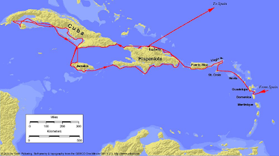
Map of second voyage (Wikipedia) Click to enlarge.

         Chanca, in a letter back to Spain, gave some idea of local problems, but he provided no further insight into the diagnosis of the epidemic disease. Disease entities as we know them had not entered the medical vocabulary. He worked hard and Columbus praised him for his efforts to aid the sick. 

Famine accompanied the epidemic. The initial crops of the Spanish colonists grew poorly, provoking the Spanish to steal from the Hispaniola native Taíno. Attempts to enslave natives or otherwise maltreat them led to hostilities that reduced the food supplies of both groups. Hunger and starvation, lowering resistance to disease, drastically drove down the population of Spaniards and natives alike. When Columbus returned to Spain in early 1494, over half of the original 1500 colonists and untold numbers of Taíno had perished. Reports of illness with superficial ulcers appeared, perhaps the first recognition of syphilis, considered a “new world disease.” The surgeon of the previous trip, Maestro Juan, and the fifteen men left with him had all perished, probably killed by local natives. 

Columbus searched for spices as well as gold. He and Dr. Chanca identified plants they thought were varieties of aloe, pepper, and cinnamon. They were incorrect, though some plants proved useful as medicines. The spice they labeled “cloves” was allspice, so named because its odor suggested a mixture of clove, cinnamon, and nutmeg. And they found tobacco.

By the 1500s, colonizers and adventurers were arriving in larger numbers, both on the islands and the mainland. Various febrile diseases, probably including malaria, were now common, though no diagnoses familiar to us appear. Smallpox arrived in the Caribbean in 1518. The native population, never exposed to it and already in severe decline, suffered severely while the Spanish population, maintained only through new immigration, had fewer losses. In 1520, smallpox reached Mexico, imported by an expedition sent

Hernán Cortés, 1525 portrait
(Wikipedia)

from Cuba to arrest Hernán Cortés for disobeying orders. Cortés had attacked Tenochtitlán (now Mexico City) but had been forced by Aztec forces to withdraw. He was licking his wounds when smallpox intervened and devastated the victorious Aztecs, reducing their numbers and killing their leader. Cortés took the depopulated city back, another example of disease affecting history. Smallpox went on to devastate Central and South America, ravaging whole populations. 


Smallpox in early America, from Florentine Codex 
(Wikipedia)

The first documented epidemic of measles broke out in 1532, though reports suggestive of its appearance surfaced as early as 1529. Measles, highly infectious, swept through the colonies rapidly, causing many more deaths. In all the outbreaks of disease mentioned, the combination of Spanish disruption in the daily life of native Americans, slavery, work conditions, and the falloff in the ability to farm and prepare food, produced widespread famine and lowered resistance to disease, all contributing to the terrible mortality.

More epidemics plagued the Spanish possessions, though their nature is often obscure. Two deadly epidemics, in 1545 and 1576, of a new disorder, known in Nahuatl as cocoliztli, killed up to 80% and 50% of the native population respectively. Symptoms included nosebleeds, hemorrhagic phenomena, diarrhea, and fever. Recent studies suggest they might have been due to a virus related to today’s arenavirus or hantavirus (“four corners virus”) species, carried in rodents. Both outbreaks followed periods of severe drought.

Much is unknown about the early medical problems of the Spanish New World and will probably remain so. But it is clear that epidemics and death flourish in times of food shortage and/or severe social disruption, even today.


SOURCES:


Cook, N D, Born to Die: Disease and New World Conquest. 1998, Cambridge Univ Press.


Gonzalez, J P H, “En Torno a Una Biografía del Primer Médico de América Diego Álvarez Chanca (circa 1450 – post 1515).” 2012; Anuario de Estudios Atlánticos 58: 29-49.

De Ybarra, A M F, “ A Forgotten Worthy, Dr. Diego Álvarez Chanca, of Seville, Spain.” 1906; JAMA 47 (13): 1013 17.

Acuna-Soto, R et al, “Megadrought and Megadeath in 16th Century Mexico.” 2002; Rev Biomed 13: 289-92. (also in Emerging Infectious Diseases 8 (4), 2002, available at: https://wwwnc.cdc.gov/eid/article/8/4/01-0175_article)

Griffinhagen, G B, “The Materia Medica of Christopher Columbus.” 1992; Pharmacy in History34(3): 131-45.

A full index of past essays is available at: 

https://museumofmedicalhistory.org/j-gordon-frierson%2C-md


Monday, April 15, 2024

 CHOLERA BEFORE ROBERT KOCH


         Cholera outbreaks today are confined to areas of famine, war, or other sites of public health disarray. Though an old disease in India, it is a relatively new disease from a global viewpoint. The first recorded pandemic, that of 1817, marched from India eastward through Asia and westward to Persia and the Caucasus, but spared Europe.

         In the next pandemic, beginning in 1829, cholera reached Russia, swept down through Europe, and traveled across the Atlantic to the United States, Canada, and South America, killing hundreds of thousands during its march. Western medical skills were powerless. Arguments raged over whether it was contagious or came from

French drawing, cholera (Wikipedia)

miasmas and/or foul air, and how to treat it. Quarantines of various types were debated and instituted irregularly. Galenic ideas, especially those involving corrupted humors to explain symptoms, still prevailed in medical thought. Accordingly, for treatment medical opinion favored measures to rid the body of corrupting material. 

The losses of fluid in cholera are dramatic. Liters can be passed in liquid stools in a few hours, exacerbated by vomiting. Records describe patients as cadaveric, with dry, cold, grayish skin, near-absent pulse, and inaudible heart sounds.  Blood from a vein or artery, if it came at all, resembled tar. Claude Bernard even suggested that survivors passed through a form of suspended animation similar to hibernation. It is obvious today that replacement of fluid is essential, but that was not so in the 1830s. Bleeding and purging were highly recommended treatments. Since blood was hard to extract, doctors often resorted to an artery to obtain small amounts of tarry material. Emetics, purgatives like calomel, cupping, and blistering were also part of regimens that did more harm than good.

Investigators in Moscow in 1830 were among the first to discover the high hematocrit and low water content of blood in cholera patients. Scattered reports of injecting water or saline solutions intravenously appeared, but results were disappointing. Inadequate amounts of fluid, injection of air bubbles, and bacterial contaminants all contributed to poor results, delaying intravenous fluid therapy until the twentieth century. Attempts to suppress bowel movements with opiates also met with little success.

A third pandemic lasted from 1845 to 1856. By then, contaminated water was under suspicion. John Snow’s surmise of a “cholera poison” in the water from a certain London water company and from a particular well resulted in removing the handle from the

John Snow (Wikipedia)

Broad Street pump in 1854. But the epidemic was already subsiding and the medical world paid relatively little attention. The “discovery” of the causative agent by an Italian microscopist also brought little reaction.

Filippo Pacini, son of a cobbler, received his medical education in Pisa. As a student, using a rudimentary microscope, he was the first to describe the tiny “Pacini” tactile dermal corpuscles, work later confirmed. His proficiency with the microscope catapulted him to the professorship of anatomy at the University of Florence. During the same pandemic that struck Snow’s London, Pacini studied cases in Florence using his microscope. He

Filippo Pacini (Wellcome Library)

noted disruptions of the intestinal mucosa and myriads of comma-shaped bacteria in the intestinal fluid. He published his findings in 1854 in an Italian journal, suggesting that the organisms caused the disease. He sent copies of his findings to the French Académie des Sciences, to Virchow, Henle, and Max von Pettenkofer in Germany. Reports also went to authorities in England, and to the Medical Society of London, where Snow was a member. Snow and Pacini never met, and it is not clear if Snow knew of Pacini’s work. Snow died in 1858 and a brief obituary in Lancet did not mention his work on cholera epidemiology.

Cholera struck Florence again in 1865. This time Pacini’s writings attracted the attention of William Farr, the British Registrar-General, who described Pacini’s findings in a report of 1868, including his insight that excess fluid loss was the cause of vascular collapse. Pacini recommended fluid replacement and developed complicated mathematical models to measure the fluid balance. Pacini’s writings, however, did not appreciably affect general medical opinion. It was too early. Lister’s first article on bacteria in wound infections appeared in 1867, was received skeptically, and Robert Koch first proved the bacterial cause of a disease (anthrax) only in 1874.

Max von Pettenkofer, the great German pioneer in public health

Max von Pettenkofer (Wikipedia)

science, explored cholera extensively. Having studied Snow’s work, he investigated water provisions in Munich and concluded that Munich’s water supply was not the problem. He went on to develop a cholera theory that required three factors to cause disease: the presence of live organisms (not yet discovered), proper soil conditions where the organisms can develop, and a susceptible host.

Finally, in 1884, Robert Koch “discovered” again the offending

Robert Koch's Egyptian team. Koch is 3rd from right. (Wikipedia)

organism during a trip to Egypt. He does not seem to have known about Pacini’s work. As the leading bacteriologist of the time, Koch was able to culture the vibrio and study its properties, techniques unavailable to Pacini. Though Pettenkofer accepted Koch’s findings, he was so assured of his soil development theory that he swallowed a pure culture of cholera vibrio, sustaining only mild diarrhea. A couple of his students who tried it, though, suffered from severe cholera. 

In India, David D. Cunningham, an army doctor in charge of cholera research, was a follower of Pettenkofer and continued to

David Cunningham (Wikipedia)

resist Koch’s thesis of direct transmission for years. But accumulating evidence overcame the last doubters and solidified the modern view of cholera transmission, pathology, and treatment. Today the disease is primarily a feature of a breakdown in water supplies and hygiene.


SOURCES:


Isaacs, J D, “D D Cunningham and the Aetiology of Cholera in British India, 1869-1897.” Medical History 1998; 42: 279-305.


Carboni, G P, “The Enigma of Pacini’s Vibrio cholerae Discovery.” J Medical Microbiology 2021; 70 (11): 1-7.


Pettenkofer, M, “Nine Propositions Bearing on the Aetiology and Prophylaxis of Cholera, Deduced from the Official Reports of the Cholera Epidemic in East India and North America.” Indian Medical Gazette 1877; April 2, May 1, July 2, and  August 1.


Howard-Jones, N, “Cholera Therapy in the Nineteenth Century.” J History of Medicine 1972; 27: 373-95.


Pollitzer, R, Cholera 1959; WHO, Geneva.


Evans, A S, “Pettenkofer Revisited: The Life and Contributions of Max Pettenkofer (1818-1901).Yale J Biol Med 1973; 46: 161-76.


Vinten-Johansen, P, et al, Cholera, Chloroform, and the Science of Medicine: A Life of john Snow. 2003; Oxford Univ Press. 


A full index of past essays is available at: 

 https://museumofmedicalhistory.org/j-gordon-frierson%2C-md


Friday, March 15, 2024

 The “Autopsy” of St. Ignacius de Loyola and the 

Anatomists Who Performed It


       On July 31, 1556, Ignatius de Loyola limped into Rome to die. The limp stemmed from a cannonball injury to his right leg sustained during his youthful days as a soldier. During convalescence Ignatius, born Íñigo López de Oñaz y Loyola in the Basque village of Loyola, experienced a spiritual conversion. After a pilgrimage to Jerusalem

Saint Igantius of Loyola (Wikipedia)

followed by studies of theology in Spain and Paris, he and colleagues in Paris formed the Society of Jesus. He expired in Rome after a long illness.

 To avoid decomposition of the body in Rome’s July heat, embalming (the custom for important religious figures) was undertaken quickly. Realdo Colombo, a famous anatomist and at that time a physician in the papal household, was assigned to open the body. Juan Valverde, a prominent Spanish anatomist, assisted him (Valverde, like Ignatius, was Basque.) Who were these anatomists and did the future saint undergo an autopsy?

         Realdo Colombo, the son of an apothecary, was born in Cremona, Italy, around 1515. After an apprenticeship with a Venetian surgeon, he moved to Padua to study medicine. His anatomy professor, Andrés Vesalius, noticing his anatomical skills, appointed him as his assistant. After a dispute with Vesalius, Colombo taught at

Realdo Colombo (Wikipedia)

Pisa and then moved to Rome where he taught at the papal university, the Sapienza, and served as a physician in the papal court. He began work on his own anatomy text, De Re Anatomica, that saw publication shortly after his death in 1559. In it he corrected some of Vesalius’ anatomical statements and, more importantly, described the pulmonary circulation and the proper function of the heart valves. William Harvey studied the work intensely, crediting it with helping him unravel the scheme of circulation in 1626. Colombo had asked Michelangelo to illustrate his planned anatomy text, but the latter was too old and died soon after.

Juan Valverde de Amusco, after obtaining a university degree in Spain, studied medicine under Vesalius and Colombo in Padua and followed Colombo to Pisa, pursuing anatomy studies. Valverde, like Colombo, came to Rome where he studied under Bartolomeo

Juan Valverde (Wellcome Library)

Eustachi, professor of medicine at the Sapienza. Valverde later taught anatomy at the Hospital Espíritu Santo, which boasted an anatomical theater. Cardinal Juan Álvarez de Toledo chose him as his personal physician, and both Valverde and Colombo moved in prominent social and scientific circles in Rome. Valverde worked closely with Colombo while investigating the pulmonary circulation. 

Valverde’s move to Rome from Spain was not unusual. The Spanish population in Rome had been growing, partly due to the election of two Spanish popes (Callixtus III, 1455-58 and Alexander VI, 1492-1503). The educational reforms of Pope Julius III (1550-55) had enhanced Roman medical education,

Bartolomeo Eustachio (Wikipedia)

reformed medical licensing, and encouraged the formation of new hospitals. In Spain, medicine was less advanced and bodies for dissection were harder to obtain. Additionally, the majority of physicians in Spain had been Jewish and the Spanish expulsion of the Jews in 1492 had driven many to Rome and elsewhere. Whether Valverde had Jewish origins is uncertain. 

Valverde published an anatomy textHistoria de la Composición del Cuerpo Humano in 1556, three years before Colombo’s work. It was well illustrated but most of the illustrations, attributed to Gaspar Becerra, who trained in Michelangelo’s workshop, are copies of those in Vesalius’ text, though smaller in size. The book includes corrections from Vesalius’ original Fabrica (which angered Vesalius), and some original findings that include a correct rendition of the pulmonary circulation, though still with Galenic overtones. The main advantage of

Striking illustration from Valverde's Historia
(Wellcome Library)

Valverde’s book is the readability and clarity of text, making it popular with medical students and physicians. The book was published in Spanish (many Spanish surgeons did not know Latin) but eventually appeared in Italian, Dutch, and Latin translations during some sixteen editions. It was one of the most widely used anatomy texts in Europe at the time. 

Colombo recorded in De Re Anatomica that inside Ignatius he found stones in the kidneys, lungs, liver, and portal vein and that the stomach and intestines were empty, the latter finding attributed to Ignatius’ frequent fasting. The dissection has often been characterized as an autopsy of Ignatius, but Colombo was there to embalm him and did not speculate on a cause of death. The nature of the “stones” is uncertain and may have alluded to gallstones, urinary tract stones, phleboliths, or some combination.

Actual autopsies were, however, not so rare at the time. Leonardo da Vinci had written in the latter 1400s, “…and I did an autopsy on him to see the cause of such a quiet death.” Antonio

Antonio Benivieni (Wikipedia)

Benivieni, a Florentine physician and contemporary of da Vinci, penned the first known book of autopsies, published posthumously in 1507. In it Benivieni describes 110 cases with postmortem findings. Normal anatomy was not fully understood at the time, not to mention abnormal anatomy, and Benivieni was steeped in Galenic medicine, a doctrine that explained health and disease in terms of the balance of humors. Thus, Benivieni did not always relate autopsy findings with the clinical picture. By Colombo’s time autopsies, still brief by modern standards, were more common. 

St. Ignatius’ death occurred at an important moment when post-Vesalian objective anatomy investigations were eroding the ancient Galenic framework of medical thought. The work by the Spanish anatomist, Valverde, was an important and often overlooked contribution. And findings at autopsy gradually reshaped concepts of disease though it was not until the great work of Morgagni in 1761, De sedibus et Causis Morborum, correlating the clinical picture with autopsy findings, that modern pathology began to take shape.


Sources:

King, L S and Meehan, M C, “A History of the Autopsy.” Amer J Pathol 1973; 73 (2): 513-44.


Andretta, E, “Juan Valverde, or Building a ‘Spanish Anatomy’ in 16th Century Rome.” Working paper, European University Institute, Max Weber Programme, 2009. https://cadmus.eui.eu/bitstream/handle/1814/12094/MWP_2009_20.pdf?sequence=1


Rosenman, L D, “Facts and Fiction: The Death of Saint Ignatius of Loyola.” Surgery, 1996; 119 (1): 56-60.


Hernandez-Mansilla, J M, “Autopsia, embalsamamiento y signos de santidad en el Cuerpo de Ignacio de Loyola.” Rev de Ciencias de las Religiones 2016; 21: 79-91.


Fye, W B, “Realdo Colombo.” Clin Cardiol 2002; 25: 135-37.

Arráez-Aybar, L, et al, “Juan Valverde de Amusco: Pioneering the Transfer of Post-Vesalian Anatomy.” Anatomia 2023; 2: 450-71. 

Burgos Lázaro, R, et al, “Juan valverde de Amusco en la Medicina del Renacimiento Español.” Anales RANM 2021; 138 (1): 82-91.

Singer, C.(translator), de Abditis Nonnullis ac Mirandis Morborum

et Sanationum Causis by Antonio Benivieni. 1954; Charles C Thomas.


Coppola, E D, “The Discovery of the Pulmonary Circulation: A New Approach.” Bull Hist Med1957; 31 (1): 44-77.


A full index of past essays is available at: https://museumofmedicalhistory.org/j-gordon-frierson%2C-md


Wednesday, February 14, 2024

                            THE DISEASE OF THE DEPOSED CHILD                                                                 

         In 1929, a 36-year-old Jamaican-born British physician, Cicely Williams, arrived by ship at the African “Gold Coast,” now known as Ghana. The inspiration for this assignment to a faraway place came from a Croatian professor of public health, Andrija Stampar, whom Williams had met on the way to work in a refugee camp near Salonika. He had shown her how to deal with with epidemics and nutritional problems at the community level using limited resources. Cicely determined to take up medicine in underserved communities as a way to confront these challenges. 

Gold Coast (Wikipedia) Click on 
image to enlarge.

 Dr. Williams, after her inspiring experience with Stampar, earned a Diploma at the London School of Hygiene and Tropical Medicine and was assigned by the British Colonial Medical Service to the town of Kumasi, in the Gold Coast. The area, formerly the land of the Ashanti empire, was now a center of cocoa production and extensive mining. 

While working in Kumasi and nearby stations, Dr. Williams encountered a clinical syndrome in young children that baffled her and did not appear in her medical texts. The afflicted children were edematous (puffed up with extra fluid), had orange hair, scaly and somewhat dry skin that was darkened in the creases, frequent diarrhea, and were mentally sluggish. Most of them died. Williams tried to obtain autopsies but was frustrated by mothers who whisked their children away before dying. After learning that the bus driver charged extra to transport a dead person, she paid the extra charge and autopsied several children. She found only an enlarged fatty liver, pallor, and edematous changes. Her first report of the disorder was in Gold Coast medical reports 1932. The local people called it “kwashiorkor.” 

Photos of kwashiorkor from Williams' first article (Wellcome Library)

Kwashiorkor was a tainted “ju-ju” word of the Ga language that locals were reluctant to pronounce. It referred to the sickness affecting a child after it is “deposed” from its privileged breastfeeding site to make way for a newborn child. The deposed child was put on a diet close or equal to an adult diet of ground maize, often laced with hot spices. The infants ate less and the reduced intake and lack of protein, sometimes mixed with parental neglect, led to what Williams determined to be a deficiency disease. She found that feeding the needy child with Nestlé’s canned, condensed milk produced a quick recovery, suggesting that protein deficiency was the main problem. Williams wrote a second report,
this one in a widely read pediatric journal, that brought the syndrome to broader attention.

Beginning of first article on kwashiorkor (Wellcome Library)

 Her second article caught the attention of the London physician, Hugh Stannus, who was an expert on pellagra, a disease due to niacin deficiency. Stannus felt that kwashiorkor was really pellagra, not a new disease. His authority held sway for a while, but eventually new evidence proved Stannus wrong. The work of Joseph Goldberger in the United States showing that pellagra was a dietary deficiency disease and the demonstration in 1937-8 that niacin alone, without a change in diet, could cure pellagra, established the difference between kwashiorkor and pellagra. 

Another champion of William’s ideas was Dr. Hugh Trowell, who worked in Uganda while Williams was in Ghana. Trowell found that medical missionaries in his area, back in the 1920s, had noticed a similar syndrome and described it in the first issue of the Kenya Medical Journal. They too had considered it a protein deficiency disease. Trowell had bowed before Stannus’ opinion but eventually joined Williams in considering it a new disease. The word “kwashiorkor” did not appear in textbooks of tropical medicine, however, until the 1950s.

Through the use of condensed milk and the encouragement of prolonged breast feeding many more children survived after abrupt weaning (though later findings showed that hypoglycemia could be a problem with condensed milk). Cicely’s work with children, mothers, and within the local communities lowered the child mortality rate, and resulted in women having fewer children. 

 A dispute between Williams and her chief led to an abrupt transfer to Malaya in 1936. Infant malnutrition in Malaya turned out to be generally due to insufficient breastfeeding from birth. Mothers, responding to European advertising and culture, had curtailed breastfeeding, feeding their infants condensed milk instead. Williams aroused controversy by campaigning to restrict the use of condensed milk and encourage milk from the breast. World War II, however, curtailed her efforts while she was incarcerated by the Japanese in the infamous Changi Prison in Singapore. After the war, still energetic, Cicely worked as head of a new Maternal and Child Health Division at WHO and was regarded as a specialist in child nutrition. After

Dr. Cicerly Williams (Wikipedia)

another stint in Malaya, she settled as a lecturer at the London School of Hygiene and Tropical Medicine. Her lectures and writings emphasized that to combat malnutrition it was necessary to investigate the social and behavioral causes, such as she had seen in Malaya, in addition to correcting the diet. She died in 1992, at the age of 98. 

Andrija Stampar, the public health physician who inspired Williams, was active in the League of Nations, and later presided over the conference convened to draft the constitution of the World Health Organization, where he served for many years.

The word kwashiorkor in textbooks now appears as part of a spectrum of nutritional disorders under the category of “Protein-calorie-deficiency” or “protein-energy-deficiency” disorders. They include kwashiorkor and marasmus, the latter characterized by severe wasting, absence of fat, and loose skin. Research work continues, as evidenced by finding distinct intestinal microbiomes in healthy and kwashiorkor-affected children. Cicely Williams’ work on nutrition lives on, as does her decades-long devotion to community maternal-child care.


A full index of past essays is available at: https://museumofmedicalhistory.org/j-gordon-frierson%2C-md


SOURCES:


     Williams, C, “Deficiency Disease in Infants: A Report by Miss C.D. Williams, Woman Medical Officer, Princess Marie Louise Hospital, Accra. Report of Medical Department, Gold Coast Colony, 1931-32. 1932; pp 93-99. 

     Williams, C D, “A Nutritional Disease of Childhood Associated with a Maize Diet.” Arch Dis Childhood. 1933; 8: 423-33.

     Craddock, S, Retired Except on Demand: The Life of Dr. Cicely Williams. 1983; Green College, Oxford.

     Trowell, H. “The Beginning of the Kwashiorkor Story inn Africa.” Central African Journal of Medicine 1975; 21(1): 1-5.

     Stanton, J, “Listening to Ga: Cicely Williams’ Discovery of Kwashiorkor on the Gold Coast.” Clio Med. 2001; 61: 149-71.

     Goodall, J, “Malnutrition and the Family: Deprivation in Kwashiorkor.”  Proc Nutrit Science. 1979; 38: 17-27. 

     Trowell, H, “Kwashiorkor, 1. Nutritional Background, History, Distribution, and Incidence.” Brit Med J.  Oct 11, 1952, 796-8.