Thursday, September 16, 2021

 A SPECIAL NOBEL PRIZE


         There is a story that the 1930 winner of the Nobel Prize in medicine and physiology first learned of his award from a Swedish journalist. After hearing the journalist, he asked what the prize was for. That was not false modesty; it was because the recipient, Karl Landsteiner, had made so many discoveries that he had to speculate on the winning one. The prize was, in fact, for the determination of the blood groups, a breakthrough that made possible the whole field of transfusions. True or not, the anecdote illustrates the wide range of Landsteiner’s achievements.

Landsteiner was born into a Jewish family in Vienna in 1868. His father, a prominent journalist, died when Karl was six. His

Karl Landsteiner (Wikipedia)

mother saw him through a gymnasium (university prep school) education, after which he entered the University of Vienna Medical School at age 17, graduating in 1891. Interested in biochemistry, he spent 5 years with prominent chemists, including Emil Fischer, and soon after learned bacteriology as assistant to Anton Weichselbaum, who had discovered the meningococcus as a cause of meningitis. He continued to work in Vienna until 1919, rising to Professor of Pathologic Anatomy at the University of Vienna and director of laboratories at the Wilhelmina Hospital. During this time his scientific output was prodigious.

In the infectious diseases, he and Ernest Finger (chief of Dermatology and Venereal Diseases) transmitted syphilis to monkeys (though preceded by Metchnikoff and Roux). On his own, Landsteiner discovered the value of dark field illumination in identifying the spirochete and found that extracts of beef heart improved the Wasserman reaction. Both were discoveries widely used. 

In 1908, during a polio epidemic, he and an assistant were the first to transmit the disease to monkeys. Landsteiner, with

 Constantin Levaditi (Wikipedia)

Constantin Levaditi, a Romanian-born scientist, established that the poliovirus was ultrafiltrable and that it was recoverable from the pharynx, salivary glands, tonsils, and in lymph nodes draining the intestines, proving that polio was a systemic infection. Landsteiner found that convalescent serum inactivated the virus and even created a vaccine. The vaccine failed to prevent disease in monkeys, though, and he abandoned the project. Landsteiner also described the pancreatic findings in what later became known as cystic fibrosis.

  
 Busts mounted in Polio Hall  of Fame, Warm Springs, GA. Landsteiner is fourth from left, 
Franklin Roosevelt is second from right. (Wikipedia)

     Regarding his Nobel Prize work, Landsteiner was long interested in blood transfusion reactions. Familiar with the phenomena of agglutination and lysis of red blood cells, he mixed serum from different donors with washed red cells. The resultant agglutinations distinguished 3 different blood groups, the fourth one discovered by his assistants soon after. This opened the door to transfusion services, especially after the introduction of the anticoagulant, citric acid. Landsteiner later uncovered other blood antigens and, finally, in 1940, he and Philip Levine discovered the Rh factor, which led to the unraveling of the perplexing problem of jaundice of the newborn. 


Diagram of Blood Groups (Wikipedia)

Landsteiner’s life was not always smooth. His mother died in 1908, a traumatic event for a man so close to her (he kept her death mask on his bedroom wall until his own death). Both he and his mother had converted to Catholicism years earlier, not an unusual practice at the time and possibly seen as an aid to his career. Though a tireless and determined worker, socially he was withdrawn and made few friends. Eight years after the death of his mother, and during WWI, he married the daughter of the verger of a Greek Orthodox Church, Helene Wlasto, who agreed to give up her Orthodox connection. A year later, 1917, a son, Ernest Karl, was born.

World War One was hard on Austria. Vienna’s glittering fin-de siècle life had disappeared and its inhabitants were barely surviving. Facilities (and salaries) deteriorated at the University and food was scarce. Landsteiner bought a goat to provide milk for his son as he watched destitute locals, desperate for firewood, cut down trees around his property. To feed his family, he moved to a job as pathologist at a Catholic hospital at The Hague. Despite the heavy workload and poor facilities, he managed to publish 12 papers. His difficulties drifted up to

 Simon Flexner (Wikipedia)
 Simon Flexner at the Rockefeller Institute in New York and, aware of his work, Flexner promptly offered him a position. 

The three Landsteiners started New York life in an apartment above a butcher shop, awakened often by the noise of passing trolleys, though he soon found a small house on Nantucket for quiet weekends. At the Institute, Landsteiner continued his impressive scientific output, concentrating on problems in immunology and serology. His work culminated in the publication of The Specificity of Serological Reactions, a landmark work in a field he had largely pioneered. He considered the work so important that, in another version of the Nobel Prize story, he thought the prize might be for his immunology research. He later collaborated with the chemist, Linus Pauling, on a new edition of his book that was published posthumously by his son, Ernest Karl, a surgeon in Boston. 

Landsteiner’s shyness and aversion to publicity caused him anxiety at the Nobel ceremonies. So much so, that when he was asked to speak at a dinner, Landsteiner deferred to that year's winner of the Prize in literature, Sinclair Lewis, who praised him as “in a thousand cases the master of death.”

Karl Landsteiner worked into his seventies, succumbing to a heart attack in 1943. Cancer took his wife six months later. Peyton Rous, a friend since his arrival in New York, wrote in an obituary appreciation that Landsteiner was “as pure a medical scientist as his material would allow… He believed that facts would make their own way and he left them to do so…” A fitting tribute. 


SOURCES:

Bendiner, E. “Karl Landsteiner: Dissector of the Blood.” Hospital Practice March 30, 1991, pp 93-104.

Gottlieb, A M, “Karl Landsteiner, the Melancholy Genius: His Time and His Colleagues.” 1998; Transfusion Medicine Reviews 12 (1): 18-27.

Goldman, A S and Schmalstieg, F C, “Karl Otto Landsteiner (1868-1943). Physician-biochemist-immunologist.” 2019; J Med Biography 27 (2) 67-75.

Bayne-Jones, S, “Dr. Karl Landsteiner: Nobel Prize Laureate in Medicine, 1930.” 1931; Science 73 (1901): 599-604.

Rous, P. “Karl Landsteiner.” In “Obituary Notices: Fellows of the Royal Society.” 1947; Proceedings of the Royal Society 5: 295-324.

Paul, J R, A History of Poliomyelitis. Chapter 11. 1971, Yale University Press.


Monday, August 16, 2021

  BURN DOWN THE QUARANTINE STATION!


         In the 1850s ships disgorged over two million immigrants into New York City, packing crowded tenement buildings to the limit. Cargo arrived too, much of it from the Caribbean where yellow fever was endemic. Frequently vessels carried epidemic diseases, of which four triggered a quarantine: smallpox, yellow fever, cholera, and typhus. Before the discoveries of bacteriology and the role of insects in disease transmission, informed opinion cited miasmas, polluted air, and fermenting ship holds as sources of these maladies. To protect the public New York had erected, in 1799, a quarantine station on Staten Island, about five miles from lower Manhattan and quite close to New Jersey. 

Map of Staten Island in 1858. X marks quarantine station. Brooklyn is to the right and Manhattan is 5 miles north. (Wikipedia and N.Y. Public Library digital collection)

Over the years, the station enlarged and played frequent host to the four diseases mentioned. Surrounding the station was a six-foot-high brick wall, sealing it from the surrounding village of Tompkinsville, while inside there were as many as 1500 people in quarantine during years of peak immigration (1840s and 1850s). Numerous personnel lived and worked within, including the medical director, a few physicians, nurses, orderlies, and various hospital employees. Resident boatmen rowed in the immigrants and stevedores brought in cargo from infected ships. 


View of station, 1858. White building with cupola behind the pier is St. Nicolas Hospital, for cabin
class passengers and VIPs only. Vegetable garden is on slope. (Wikipedia and N.Y. Public Library)


Duty was considered hazardous. Four physicians died of typhus in 1851 and another in 1856. Thirty-three workers came down with yellow fever in 1856. Needless to say, the residents of the surrounding towns feared this fountain of pestilence next door. Though much of Staten Island was thinly-populated farmland, several villages and towns along the eastern shore, near the station, suffered yellow fever outbreaks over the years. Whether they originated from the quarantine station or not, the locals saw the quarantine station as the source. They were particularly angry at the staff, who, in defiance of rules, often visited the local communities for shopping or pleasure. 

Staten Islanders had actually resisted the quarantine station from the start, but New York State, heedless, exerted “the right of eminent domain” over 30 acres of land and built the station. Small outbreaks of cholera and yellow fever harassed the communities intermittently and large outbreaks of yellow fever attacked in 1848 and 1856. After the 1848 flare-up, the New York legislature agreed to move the station to Sandy Hook, New Jersey, but New Jersey objected, shippers did not want a change, and the matter drifted. After the 1856 episode, the N. Y. Legislature authorized a station on the other side of Staten Island but the residents there burned it down twice during construction. Meanwhile, at the original station, the locals put up their own fence and insisted on a Harbor Police force to keep station personnel from wandering out. Threats and counterthreats mounted. Finally, on September 1, 1858, the Tompkinsville Board of Health met and recommended that citizens protect themselves by “abating this abominable nuisance without delay.”

That very night men crept through a hole in the station’s wall, stuffed straw mattresses in strategic sites in both hospital and residential buildings and set them on fire. The Fire Department arrived, but did nothing, claiming that their hoses had been cut. The Harbor Police were overwhelmed. The mob’s ringleader agreed to spare a building to house the patients in exchange for releasing prisoners taken. The mob grew to several hundred, opened another hole in the wall with a battering ram, and incinerated more buildings. Some patients were placed in the spared Female Hospital, others remained outdoors. 


Newspaper sketch of burning the quarantine station. (Wikipedia and N.Y. Public Library)


The next day the N.Y. Mayor and the Police Chief promised police backup, but none arrived. Meanwhile Richmond County, where the station was, had a stormy meeting during which angered residents resolved to finish the job. Station staff moved their furniture to the street and watched that night as the mob burned down all the remaining buildings, including the Female Hospital and the pier. Patients from the hospital, moved outside, were stuck between two burning buildings and had to be cooled down with buckets of water. Other patients and staff sat in the rain that fell during the night.

The New York Times castigated the Staten Islanders as lawless and selfish, while the New York Herald, Horace Greely’s paper, was more sympathetic, criticizing the New York authorities for not heeding the Staten Islanders’ repeated pleas for relief. To maintain peace, this time over 100 policemen and the State militia arrived  and tents were supplied to house the sick. Several of the mob were jailed for breaking quarantine regulations but Cornelius Vanderbilt, born on Staten Island, bailed them out. Two ringleaders were prosecuted by the State. Their defense, buttressed by witnesses, was that the station personnel had repeatedly broken rules by wandering into the neighboring communities, spreading disease, and that the mob had acted in self-defense and were seeking “freedom from State tyranny.” The judge ruled in their favor, brushing aside the charges of violence and property destruction. The judge, probably not impartial, owned property on the island and his brother-in-law had died of yellow fever.

The quarantine station moved temporarily to a large vessel, the Florence Nightingale, anchored further out in the harbor. Meanwhile, two islands, Hoffman and Swinburne Islands, were created artificially (opened in 1866) to house quarantined passengers, one island for the sick, and one for those who were well but exposed to disease. 


Quarantine hospital on Swinburne Island, 1879 (Wikipedia and 
New York Public Library)

The moral of the story? Quarantine is fine, but "not in my back yard." Using islands, as New York finally did, frequently solved the problem. Another example was the choice of Angel Island for the quarantine station at San Francisco.


SOURCES:

Garrison, F, “The Destruction of the Quarantine Station on Staten Island in 1858.” 1926; Bull N Y Acad Med 2 (1): 1-5.

N Y Times, Sept. 2 through 5, 1858.

N Y Herald, Sept 2 through 5, 1858.

Stephenson, K, “The Quarantine War: The Burning of the New York Marine Hospital in 1858.” 2004; Pub Health Reports 119 (Jan-Feb): 79-92.


Friday, July 16, 2021

 THE GUILLAIN-BARRÉ SYNDROME:

ORIGINS


     The FDA recently altered the labeling on the J&J Covid vaccine, warning of a small risk of Guillain-Barré syndrome in recipients. GBS, as it is sometimes called, has an interesting history. 

     Guillain, Barré, and Strohl (yes, three authors) first described the syndrome in two French soldiers during the First World War. The soldiers suffered from an ascending paralysis extending up to their arms, and dysesthesias, from which they recovered almost completely. Lumbar punctures were done on both, the fluid showing a high protein content and no polymorphonuclear (inflammatory) cells, a finding the authors said was distinctive. Lumbar puncture was relatively new at the time. Heinrich Quincke and Walter Wynter

Heinrich Quincke (Wikipedia)

had almost simultaneously published the technique in 1891. Both physicians aimed to relieve elevated pressure and not to study the fluid.

     The first of the 3 GBS authors, Georges Guillain, trained in neurology under Pierre Marie at the famous Salpêtrière Hospital in Paris. He was working his way up the academic ladder when the war broke out and promptly volunteered for service. He served in various field units, eventually becoming chief physician of the neurology section of the Sixth

Georges Guillain (Wikipedia)

Army. Jean-Alexandre Barré trained under Joseph Babinski and Pierre Marie. In the war he also worked in various field stations, eventually heading a military neurology center in the Eastern Region. 

     Neurologic and brain injuries were especially common in the early years of WWI because of poorly designed French helmets that provided inadequate protection. Both La Salpêtrière and La Pitié hospitals militarized their neurology departments to handle the flood of patients. At the former, Pierre Marie noted that “War injuries have shown us a different set of facts: lesions of the cortex, with more or less exclusion of white

Jean-Alexandre Barré (Wikipedia)

matter. Consequently, this new pathology is infinitely closer to the data of experimental physiology than was the old cerebral pathology.” At La Pitié Hospital, Joseph Babinski ran the military neurology unit. Guillain and Barré enjoyed a fruitful collaboration, publishing important papers on injuries to the brain and the spinal cord and delineating indications for surgery.

     The third author of the original paper, André Strohl, had trained in physics and medicine. Strohl performed electromyograms on the calf muscles of the two

André Strohl (Wikipedia)

patients, showing a weakening or absence of the reflex response and a delay in response. It was an early use of the technique. After the war, Strohl worked as professor of physiologic medicine in Algiers and later professor of physical medicine at the University of Paris. He was elected to the Académie de Médecine at the age of 35, an unusual honor. He wrote several books on nerve and muscle physiology, on nuclear isotopes, and a text on physical medicine. 

     Of course, other physicians had published cases of ascending paralysis before the war. Jean Baptiste Octave Landry, the best-known, had reported on acute ascending paralysis (10 cases) in 1859, before the age of lumbar puncture. Other reports followed, and a few earlier ones came to light. The term “Guillain-Barré syndrome” was first used by Barré at a neurology conference in 1927.  Guillain and Barré maintained that their syndrome (they actually called it “notre syndrome”) was distinct, emphasizing the favorable recovery and the findings in the spinal fluid. Why the name Strohl was dropped from “our syndrome” by Guillain and Barré, was never quite clear. Various reasons have been suggested: his German name and his origin from Alsace and the fact that he was not a neurologist. Only one month after the original Guillain-Barré-Strohl report, Pierre Marie and Charles Chatelin published 3 more cases in soldiers, acknowledging the earlier report by G, B, and S. With slightly different timing, might this have been dubbed the “Marie-Chatelin” syndrome? 

Salpêtrière Hospital, about 1660, after conversion from gunpowder factory (saltpeter was used to make
gunpowder). Image by Jean-Pol GRANDMONT of engraving by Adam Pérelle (Wikipedia).

     Over the years, many more cases of ascending paralysis came to light, presenting with varying degrees of paralysis and recovery and not always with “typical” spinal fluid findings. Many were fatal. Guillain and Barré resisted the trend to broaden the spectrum of “their syndrome” but eventually they had to yield. The GBS syndrome now includes a wide range of presentations.

     The range is wide enough, in fact, that a group of 5 authors recently wondered if Franklin D. Roosevelt might have suffered from GBS instead of polio. In early August 1921, Roosevelt, a 39-year-old practicing lawyer, was vacationing on Campobello Island, New Brunswick. After a day of vigorous activity, he developed aches and sensitivity to touch in his legs. His legs weakened and over a few days were completely paralyzed. Eventually the family consulted Robert W. Lovett, professor of orthopedics at Harvard, who had organized the first polio clinic at Boston Children’s Hospital. He diagnosed poliomyelitis and recommended a spinal tap, though it appears not to have been done. Roosevelt entered Presbyterian Hospital in New York under the care of Dr. George Draper, a classmate of FDR’s at Groton and Harvard, who had written a book on polio in 1917. As we all know, Roosevelt’s legs never recovered.

          Was FDR’s disease really polio? Or could it have been Guillain-Barré syndrome? To make a case for GBS, the 5 authors pointed out differences between typical polio and GBS syndromes, using a statistical analysis to conclude that GBS was the more likely diagnosis. A subsequent paper challenged that conclusion. Lovett and Draper may not have seen the G, B, and S paper of 1916, written in French. Whatever the truth, it is likely that if Roosevelt’s diagnosis had not been polio the March of Dimes, the principal driver of the first polio vaccine, would not have come into being.


SOURCES:

Waclawik, A J, “The Legacy of the Seminal Publication by Guillain, Barré, and Strohl: The History Behind the Eponym.” 2018; Wisconsin Med J 117: 160-3.


Pietrzak, K, et al, “Georges Guillain.” 2016;  J Neurol 263: 2148-9.


Green, D, “Infectious Polyneuritis and Professor André Strohl – A Historical Note.” 1962; New Engl J Med 267: 821-2.


Goldman, A S, et al, “Franklin Delano Roosevelt’s (FDR’s) (1882-1945) 1921 Neurologic Disease Revisited; the Most Likely Diagnosis Remains Guillain-Barré Syndrome.” 2015; J Med Biog 24 (4): 452-9.


Goldman, A S, et al, “What Was the Cause of Franklin Delano Roosevelt’s Paralytic Illness?” 2003; J Med Biog 11: 232-40.


Dittuno, J F, et al, “Franklin Delano Roosevelt: The Diagnosis of Poliomyelitis Revisited.” 2016; J Phys Med Rehab 8: 883-93.


Guillain, G, Barré, J-A, et Strohl, A, “Sur un Syndrome de Radiculo-Névrite avec Hyperalbuminose du Liquide Céphalo-Radicien sans Reaction Cellulaire.” 1916; Bull Memoires Société Hosp Parisser 3, v40,prt 2: 1462-70.


Walusinski, O, et al, “French Neurologists During World War One.” 2016; Frontiers Neurol Neuroscience 38: 107-118. 


Monday, June 14, 2021

 FROM SIENA TO NAPOLEON: 

A TALE OF TWO ANATOMISTS


         The lymphatic system occupies but little time in today’s anatomical studies. Hard to see, the tiny, colorless lymphatic vessels nevertheless are ubiquitous and serve important functions, as described by the anatomist who pioneered investigations into their pathways, Paolo Mascagni. Born in a village near Siena, Tuscany,

Paolo Mascagni (Wikipedia)

Paolo studied at the University of Siena. His professor of anatomy, Pietro Tabarrani, noticed his talents, appointed him as prosector, and advised him to study the lymphatic vessels. When Tabarrini died in 1780, Mascagni assumed his chair and explored the lymphatic system intensively. In 1784 the Academy of Sciences in Paris opened a competition for the best work showing lymphatic vessels. Mascagni entered two papers that arrived too late to win the prize. But the Academy, impressed, awarded him a special prize and shortly afterward he published a monumental volume on lymphatics, illustrated with 41 copper engravings. He is credited with discovering about 50% of the lymphatic vessels, showing that they originated from cavities and surfaces, that they all passed through one or more lymph nodes, and that they were separate from the arteriovenous system except at their termination at the thoracic ducts (in contrast to general belief). 

     Painstaking work was the secret to his success. Using mercury as a contrast agent, he injected it through glass tubes whose finely tapered ends were inserted into lymph vessels. The detailed drawings tell the story (see illustration).

Thoracic lymphatics, from Mascagni's Vasorum
Lymphaticorum (Hathi Trust)


     In 1797, France’s revolutionary army invaded Tuscany, an area under Austrian rule since 1737. Under the French regime, Mascagni reluctantly served as superintendent of arts, sciences, and charitable institutions, fighting against the removal of valuable property to France. When the Austrians regained control, he was branded as a collaborator, jailed, then freed after court battles. In 1801, realizing that his punishment was probably unjust, the University of Florence attracted him by combining the chairs of anatomy, physiology, and chemistry and raising his salary. In Florence he wrote a student’s anatomy text and prepared a series of life-sized wax anatomical models, a popular method for teaching before the introduction of cadaver preservation techniques.

     Mascagni passed away suddenly in 1815 of a septicemia. Two years earlier he had taken on a bright student, Francesco

Francesco Antommarchi 
(Wikipedia)

Antommarchi, as prosector. Francesco, born in Corsica, having earned a degree in medicine and a doctorate in surgery, had assisted Mascagni in the preparation of the student’s anatomy atlas. Mascagni’s heirs tasked Antommarchi with putting two unfinished works of the anatomist into publication. One, on histology, was published quickly. The other was Mascagni’s major work, a huge color atlas of anatomy. Before it was completed, however, news arrived that Napoleon, in exile on St. Helena after losing at Waterloo, needed a new physician since the Irish one assigned to him was leaving. Antommarchi, a Corsican like Napoleon and actively promoted for the job by his mother and family, was chosen as Napoleon’s new physician. When he left Florence, Antommarchi took three sets of plates with him, expecting to finish the atlas later. 

     He arrived in 1815 at St. Helena, an island owned by the British East India Company that was 122 miles from the nearest land. To prevent a second escape by the ex-emperor, six brigs (2-masted square-rigged vessels) sailed around the island day and night, five other armed vessels were kept near the capital, and 2784 military

Archibald Arnott (Wikipedia)

men kept watch on land. Napoleon, age 45 and in good health at the time, considered Antommarchi young and inexperienced and intermittently dismissed him, only to reconsider. But to satisfy Napoleon, the British brought a British army surgeon, Archibald Arnott, who had attended the wife of Napoleon’s Grand Marshall, to the island in 1821. By this time Napoleon was ill, losing weight, vomiting intermittently, and complaining of upper abdominal pains. Treatments of calomel, blistering, and the like did not help and Napoleon died on May 5, 1821. An autopsy revealed extensive stomach cancer. Dr. Arnott and Antommarchi both made death masks. 
"Death of Napoleon" by Charles de Steuben. Antommarchi is to left of Napoleon with hand on pillow, 
seated below him is General Bertrand, an old comrade, whose wife with two children are to the right.
A third child peeks behind the pillows. Dr. Arnott is the second head in from the right margin.
Napoleon's valet and a servant behind him are framed by the canopy. Click on plate to enlarge.
(Painting from Wikipedia. For labels see: https://www.napoleon.org/en/history-of-the-two-empires/articles/who-was-present-at-napoleons-death/ )

     Meanwhile, the managers of Mascagni’s estate had cancelled Antommarchi’s contract for the large atlas and assigned it to three professors at Pisa. They produced the great Anatomia universa over a period of nine years. The work is a major achievement. Its pages

Plate from Anatomia Universa
Click on pic to enlarge it. (Hathi Trust)

are roughly 3 x 2 
½ feet in size and many plates are hand colored. The figures are arranged so that, in some cases, three pages, laid together, combine to show a layer (muscles, for example) of a human in life-size. The principle signature on the plates is that of Antonio Serantoni, a known artist and engraver.

     Antommarchi, defying a court order, produced his own version of the work, using the plates in his possession. It was not successful, was technically inferior, and lacked a number of figures that appear in the Anatomia. After his publication, Antommarchi was restless. He became an inspector of hospitals in Poland, where he assisted in the 1831 uprising against Russia, but fled to Paris as the revolt failed. From there he moved to Louisiana, then Veracruz, Mexico, and finally settled in Santiago, Cuba, where his cousin owned a plantation. There he acquired a reputation as a skilled ophthalmologist, specializing in cataract surgery. He never married and died of yellow fever at the age of 57, while living in the Governor’s house.

     Mascagni’s contribution to the anatomy and function of lymphatics is a lasting one. His Anatomia, however, is now prized primarily by collectors. Modern students turn to their computer screens for 3D anatomy models.


SOURCES:

Eimas, R. “The Great Anatomy of Paolo Mascagni.” Available at: 

https://ir.uiowa.edu/bai/vol38/iss1/5/

Di Matteo et al, “Art in Science: Giovanni Paolo Mascagni and the Art of Anatomy.” Clin Orthop Relat Research (2015) 473: 783-8.

Riva, A., et al, “The Evolution of Anatomical Illustration and Wax Modelling in Italy from the 16thto Early 19th Centuries.” J of Anatomy (2010) 216: 209-222.

Wilson, J. B. “Dr. Archibald Arnott: Surgeon to the 20th Foot and Physician to Napoleon.” Brit Med J (1975) August 2, pp 293-5.

Homason, Henry D., Napoleon, The First Emperor of France: Being a Summary of the Facts Concerning the Latter Days of Dr. Francois Antomarchi, the Last Physician to his Imperial Majesty.1910, Franklin Hudson Pub Co., (The spelling of Antommarchi’s name varies)

Monday, May 17, 2021


NAPOLEON'S CHIEF MEDICAL OFFICER 

AND THE PLAGUE IN EGYPT      


     Napoleon made efforts to ensure good medical care for his troops. His chief surgeon, Dominique Larrey, is justly famous for his skills and compassion for the wounded. Less well known is Napoleon’s chief medical officer, René-Nicolas Dufriche-Desgenettes, a man of depth and courage.   

René-Nicolas Desgenettes (Wikipedia)

     Desgenettes was born in 1762 into a monied family in Rouen. After training in the classics, he switched to medicine and studied in Paris, England, and Italy, absorbing literature and art along with his medical pursuits. In Siena he befriended Paolo Mascagni, a great anatomist who mapped out the human lymphatic system for the first time. When Desgenettes eventually obtained his MD degree in Montpellier, he wrote his dissertation on the lymphatic system.

The Lymphatic vessels of the Back
(from Mascagni's Atlas, Hathi Trust)

     The French revolution was underway at that time and since Desgenettes’ wealthy background put him in danger, he sought refuge as a physician in the army. He met Napoleon by chance during a campaign near Nice and impressed the 24-year-old artillery officer with his intelligence and knowledge. Later, when Napoleon planned his invasion of Egypt, he asked Desgenettes, now a professor at the French military medical school, to be army’s chief medical officer. The chief surgeon, Dominique Larrey, worked under him.

Dominique Larrey (Wikipedia)

     Napoleon’s forces landed at Alexandria, Egypt, in July 1798, and fought their way to Cairo. The health problems encountered were almost as bad as the fighting. Dysentery sent many to the grave. Trachoma, the “Egyptian eye disease,” inflamed the eyes of almost everyone. Heat prostration and dehydration drove men to drink unfiltered water, often inhabited by tiny thread-like creatures. They were leeches that attached themselves to the pharynx, became engorged with blood, and provoked coughing, difficulty breathing, and sometimes hemorrhage. They could be detached with gargles of vinegar and saltwater or extracted with forceps. Smallpox, fevers, venereal diseases, and plague assailed the troops at various times. After fresh supplies from the sea were cut off by Admiral Nelson’s fleet, scurvy set in. 

     The cause of almost all these maladies was unknown. Desgenettes instituted regulations on hygiene, clothing, food and water, waste disposal, etc. and clamped down on prostitution, but much of the army still suffered from disease. Larrey established a camel-based ambulance service for wounded troops.


Larrey's ambulance service in Egypt. The lower portion shows the wounded man fitting into the compartment. (from Description de l'Égypte, courtesy New York Public Library)


     The disease that instilled the most terror, though, was plague. The role of fleas and rats was unknown at the time and the army treated it simply as a contagious disease. Plague first broke out in Egyptian coastal cities. Desgenettes thought that inadequate perspiration impaired recovery and encouraged wearing clothing in bed at night. To allay fear, he downplayed plague's importance but ordered the  burning of contaminated clothing and implemented isolation of patients and quarantines. 

     Napoleon soon lost his fleet to the British and learned that the Ottomans planned to move in from the northeast. Feeling trapped, he began an offensive into Syria (now Israel and Palestine) to neutralize the Ottoman front. During vicious fighting at Jaffa, plague broke out in force and sick troops soon overwhelmed the hospital set up in a Greek Orthodox monastery. Up to 15 soldiers died per day, according to Larrey.


     Napoleon believed that fear of the plague made one more likely to die from it. To allay such fear, he walked through the plague hospital, and, according to Desgenettes, spoke to “almost all the soldiers conscious enough to hear him.” In a small ward he helped to carry “the hideous corpse of a soldier whose torn uniform was soiled by the spontaneous bursting of an enormous abscessed bubo.” The action stunned the troops and boosted morale. The dramatic scene inspired a painting by Antoine-Jean Gros, a former pupil of David, finished in 1804 (see below). 

Les Pestiferés de Jaffa (Wikipedia) In a preliminary sketch Gros had depicted Napoleon carrying a dead soldier, but changed the composition to the above. Napoleon points to a bubo. Desgenettes' head is just to the right of Napoleon's. An army doctor is dying in the lower right and a robed Ottoman physician kneels near him. The monastery setting has been changed to that of a mosque.

     Napoleon ordered Roman Catholic and Armenian Christians to perform nursing duties in the plague hospital, and the Greek Orthodox to work in a hospital for the wounded. In general, though, hospital attendants were “the scum and disgrace of society,” recruited from prisons and galleys (Desgenettes). They were poorly paid but profited from the sale of clothing and effects taken from the dead or dying, despite rules to burn those articles. Napoleon had ordered any caught in such behavior to be shot, though how many were executed is unclear.

     Further north, during a siege in the ancient city of Acre, Desgenettes set up another plague hospital. Like Napoleon, he sought to reduce fear among the troops. One day, to reassure them, he plunged his lancet into a patient’s bubo and made small incisions with it in his groin and axilla, then washed himself with soap and water. Aside from mild inflammation he came to no harm. He also drank from a glass offered by a dying patient. The mood of the troops is said to have improved greatly. These attempts to show that plague was not dangerous or contagious may well have enhanced further spread, as soldiers relaxed hygienic and isolation habits. Treatments, including bleeding, blistering, emetics, and medicines were useless. 

Desgenettes inoculating himself with plague (from Chururgiens et Blessés a Través l'Histoire, 
by Cabanés, Hathi Trust)

      Napoleon was unsuccessful in subduing Acre and ordered a humiliating retreat back to Cairo. Returning through Jaffa, he faced a hospital filled with plague-infected troops, some of whom were near death and unable to make the blistering desert journey ahead. He recommended that Desgenettes administer large doses of laudanum to ease the hopeless ones into a rapid, painless death and avoid Ottoman cruelties. Desgenettes refused, though the doses were administered by someone else. Estimates are that between 15 and 50 men were dispatched this way.

     On arrival in Cairo, about 700 men had been lost to the plague, 4000 men overall.

     Napoleon evidently appreciated Desgenettes’ adherence to the Hippocratic oath. He kept him on as chief medical officer during his invasion of Russia and at Waterloo, and even made him a baron. Desgenettes acted courageously and humanely during a brutal Egyptian campaign but made no new discoveries about the diseases crippling his troops.


SOURCES:

Burleigh, N. Mirage: Napoleon’s Scientists and the Unveiling of Egypt. 2007; Harper Collins.

Herald, J. C. Bonaparte in Egypt. 1962; Harper & Row.

Richardson, R.G.  Larrey: Surgeon to Napoleon’s Imperial Guard. 1974; John Murray.

Gazel, L. Le Baron Des Genettes (1762-1837): Notes Biographiques. Thesis, 1912, Henry Paulin, Paris.

Eimas, Richard. “The Great Anatomy of Paolo Mascagni.” 1983; Books at Iowa no.38. available at: https://ir.uiowa.edu/bai/vol38/iss1/5/

Russell, T G and Russell T M. “Medicine in Egypt at the Time of Napoleon Bonaparte.” 2003; BMJ v327 (Dec 20-27): 1461-4.

Riaud, Xavier. “René-Nicolas Dufriche, baron Desgenettes (1762-1837), surgeon of the Great French Army.” The Napoleon Society of Ireland, at: http://napoleonireland.com/napoleon/academic-papers-articles/surgeon-to-the-great-army/

Kelly, C. "Medicine and the Egyptian Campaign: The Development of the Military Medical Officer during the Napoleonic Wars c. 1798-1801." 2010; Canad Bull Medical Hist 27: 321-42.

Roberts, A. Napoleon: A Life. 2014; Penguin Group.

English translation of: Larrey, D J. Memoirs of Military Surgery and Campaigns of the French Army, vol I. 1814; Joseph Cushing, Baltimore.     


Wednesday, April 14, 2021

 ANATOMY IN PHILADELPHIA AND SIAMESE TWINS


    In 1906, Franklin Mall, professor of anatomy at Johns Hopkins during the time of William Osler and William Halsted, gave a speech to the Association of American Anatomists, of which he was president. In reviewing the history of anatomy in America he said, “More than a century ago the status of anatomy in America compared favorably with that in Europe.” He signaled out the University of Pennsylvania, where anatomy instruction began in 1765. The first professor was William Shippen Jr., followed by Caspar Wistar. Wistar had studied in England and Edinburgh, wrote the first American textbook on anatomy, and provided a valuable collection of wax-injected preserved anatomic

Caspar Wistar (Wikipedia)
specimens. He was succeeded by Philip Physick, also known as “the father of American surgery,” whose interest was primarily surgical, and then by William E. Horner, who brought out new editions of Wistar’s anatomy text and founded an anatomical museum.
Last in the illustrious line of anatomy professors was Joseph Leidy, “the greatest teacher of anatomy to medical students this country has seen (Mall).” Leidy was professor of anatomy from 1853 until his death in 1891. By then the Wistar and Horner Anatomy Museum was aging. It was rescued in 1892 by Caspar Wistar’s great-nephew, Isaac Wistar, a general in the Civil War who had become wealthy in the railroad business. He built a new facility, the Wistar Institute for Anatomy and Biology. The Wistar Institute is still active today in a broad array of scientific endeavors.

     In 1874, a unique anatomic specimen became available to anatomists and surgeons in Philadelphia. The famous Siamese Twins, Chang and Eng, the source of headlines for years, had died. The Siamese Twins were born on a riverboat in southern Thailand in 1811. The mother was of mixed Chinese-Thai heritage, the father was Chinese. They were joined at the sternum with a band of tissue that in adulthood measured about five inches in length and eight inches in circumference. The twin on the right, Chang, was slighter in 

The Siamese Twins, painted by Édouard Pingret 1836 (Wikipedia)

development than Eng and had a more irritable personality. A Scottish roving businessman and a ship captain brought them to America as teenagers and exhibited them as “freaks” for several years, netting their “owners” handsome profits. After one partner cashed out and the other died, the twins exhibited themselves for several years, then settled in North Carolina, where they purchased a farm to raise tobacco, married a pair of white sisters, and fathered twenty-one children. They purchased slaves to work the farm. 

     In 1870, on a return trip from Europe by ship, while playing chess with the son of a fugitive slave, who was now president of Liberia, Chang developed a right hemiplegia. Four years later Chang developed a chest infection and died during the night. When Eng woke up and learned that his brother was dead, he too passed on quietly a few hours later.

     On hearing of their death, Joseph Pancoast, a Philadelphia surgeon, telegraphed an inquiry about performing an autopsy on the twins. Pancoast was head of the anatomy

Joseph Pancoast (Wikipedia)

department at Jefferson Medical College, had taken over the editing of Wistar and Horner’s anatomy text, and was known for his extensively illustrated surgery text. Dr. Joseph Hollingsworth, the twins’ personal physician, felt that burying the twins locally would invite grave robbers. Rumors of a reward for the body were already circulating. 

     With assent from the family, Pancoast and Harrison Allen, professor of comparative anatomy at the U of Penn, came to their home, embalmed the bodies with zinc chloride solution, and shipped them to Philadelphia for an autopsy.

     Philadelphia, a prominent medical city and for many years a leader in anatomical studies, was an appropriate choice. The city boasted five medical schools, was home to the American College of Physicians and was the site of the founding of the AMA in 1847.

Pennsylvania Hospital 1755 (Wikipedia)

Joseph Leidy, the “greatest teacher of anatomy,” chaired the above-mentioned U of Penn anatomy department. Joseph Pancoast, 
chairman of anatomy at Jefferson, was described as a gifted operator with “hands as light as floating perfume.” The great Samuel Gross was chairman of surgery at Jefferson and was to have his portrait painted the following year by Thomas Eakins  in “The Gross Clinic.” S. Weir Mitchell practiced in the city as a celebrated neurologist and novelist and the famous Mütter Museum, a gift of the surgeon Thomas Mütter, had opened. 

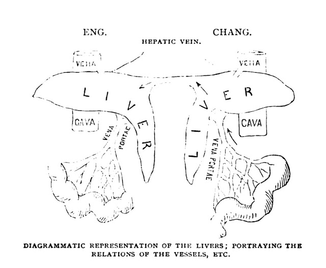
     The findings of the limited autopsy on the twins were reported during a meeting at the College of Physicians. The family permitted only the posterior aspect of the band to be opened, along with limited abdominal incisions. The major findings were that the twins shared liver tissue through the connecting canal, that their respective peritoneal membranes overlapped each other in the canal but remained separate, and that umbilical arteries terminated feebly at the entrance to the band (the umbilicus was considered shared in the center). Dye injected into the portal circulation on Chang’s side emerged on Eng’s side.

(From the article referenced below)

     During life it had become clear that the two arterial circulations and nervous systems were distinct. When Chang became intoxicated Eng did not, and if Eng ate asparagus only his urine smelled of it later. Similarly, if the center of the connecting band was pricked, they both felt pain, but not if pricked closer to their respective bodies.
  

     The autopsy satisfied the doctors’ curiosity but did not open any new doors. The bigger lesson, it seems, was that two conjoined individuals could tolerate each other for 63 years.


SOURCES:

Huang, Yunte. Inseparable: The Original Siamese Twins and their Rendezvous with American History. 2018; Liveright.

Report of the Autopsy of the Siamese Twins. (Reprinted from the Philadelphia Medical Times), 1874. Available on Hathi Trust.

Mall, F P. “On Some Points of Importance to Anatomists.” 1907; Science 25(630): 121-5.

Savitch, S L, et al. “Joseph Pancoast, MD (1805-1882): The Surgeon Who Brought Anatomy to Life.” 2020; The American Surgeon. (online only)

Rutgow, I. “American Surgical Biographies.” Surgical Clinics of North America, 1987; 67(6): 1153-80.