Monday, June 14, 2021

 FROM SIENA TO NAPOLEON: 

A TALE OF TWO ANATOMISTS


         The lymphatic system occupies but little time in today’s anatomical studies. Hard to see, the tiny, colorless lymphatic vessels nevertheless are ubiquitous and serve important functions, as described by the anatomist who pioneered investigations into their pathways, Paolo Mascagni. Born in a village near Siena, Tuscany,

Paolo Mascagni (Wikipedia)

Paolo studied at the University of Siena. His professor of anatomy, Pietro Tabarrani, noticed his talents, appointed him as prosector, and advised him to study the lymphatic vessels. When Tabarrini died in 1780, Mascagni assumed his chair and explored the lymphatic system intensively. In 1784 the Academy of Sciences in Paris opened a competition for the best work showing lymphatic vessels. Mascagni entered two papers that arrived too late to win the prize. But the Academy, impressed, awarded him a special prize and shortly afterward he published a monumental volume on lymphatics, illustrated with 41 copper engravings. He is credited with discovering about 50% of the lymphatic vessels, showing that they originated from cavities and surfaces, that they all passed through one or more lymph nodes, and that they were separate from the arteriovenous system except at their termination at the thoracic ducts (in contrast to general belief). 

     Painstaking work was the secret to his success. Using mercury as a contrast agent, he injected it through glass tubes whose finely tapered ends were inserted into lymph vessels. The detailed drawings tell the story (see illustration).

Thoracic lymphatics, from Mascagni's Vasorum
Lymphaticorum (Hathi Trust)


     In 1797, France’s revolutionary army invaded Tuscany, an area under Austrian rule since 1737. Under the French regime, Mascagni reluctantly served as superintendent of arts, sciences, and charitable institutions, fighting against the removal of valuable property to France. When the Austrians regained control, he was branded as a collaborator, jailed, then freed after court battles. In 1801, realizing that his punishment was probably unjust, the University of Florence attracted him by combining the chairs of anatomy, physiology, and chemistry and raising his salary. In Florence he wrote a student’s anatomy text and prepared a series of life-sized wax anatomical models, a popular method for teaching before the introduction of cadaver preservation techniques.

     Mascagni passed away suddenly in 1815 of a septicemia. Two years earlier he had taken on a bright student, Francesco

Francesco Antommarchi 
(Wikipedia)

Antommarchi, as prosector. Francesco, born in Corsica, having earned a degree in medicine and a doctorate in surgery, had assisted Mascagni in the preparation of the student’s anatomy atlas. Mascagni’s heirs tasked Antommarchi with putting two unfinished works of the anatomist into publication. One, on histology, was published quickly. The other was Mascagni’s major work, a huge color atlas of anatomy. Before it was completed, however, news arrived that Napoleon, in exile on St. Helena after losing at Waterloo, needed a new physician since the Irish one assigned to him was leaving. Antommarchi, a Corsican like Napoleon and actively promoted for the job by his mother and family, was chosen as Napoleon’s new physician. When he left Florence, Antommarchi took three sets of plates with him, expecting to finish the atlas later. 

     He arrived in 1815 at St. Helena, an island owned by the British East India Company that was 122 miles from the nearest land. To prevent a second escape by the ex-emperor, six brigs (2-masted square-rigged vessels) sailed around the island day and night, five other armed vessels were kept near the capital, and 2784 military

Archibald Arnott (Wikipedia)

men kept watch on land. Napoleon, age 45 and in good health at the time, considered Antommarchi young and inexperienced and intermittently dismissed him, only to reconsider. But to satisfy Napoleon, the British brought a British army surgeon, Archibald Arnott, who had attended the wife of Napoleon’s Grand Marshall, to the island in 1821. By this time Napoleon was ill, losing weight, vomiting intermittently, and complaining of upper abdominal pains. Treatments of calomel, blistering, and the like did not help and Napoleon died on May 5, 1821. An autopsy revealed extensive stomach cancer. Dr. Arnott and Antommarchi both made death masks. 
"Death of Napoleon" by Charles de Steuben. Antommarchi is to left of Napoleon with hand on pillow, 
seated below him is General Bertrand, an old comrade, whose wife with two children are to the right.
A third child peeks behind the pillows. Dr. Arnott is the second head in from the right margin.
Napoleon's valet and a servant behind him are framed by the canopy. Click on plate to enlarge.
(Painting from Wikipedia. For labels see: https://www.napoleon.org/en/history-of-the-two-empires/articles/who-was-present-at-napoleons-death/ )

     Meanwhile, the managers of Mascagni’s estate had cancelled Antommarchi’s contract for the large atlas and assigned it to three professors at Pisa. They produced the great Anatomia universa over a period of nine years. The work is a major achievement. Its pages

Plate from Anatomia Universa
Click on pic to enlarge it. (Hathi Trust)

are roughly 3 x 2 
½ feet in size and many plates are hand colored. The figures are arranged so that, in some cases, three pages, laid together, combine to show a layer (muscles, for example) of a human in life-size. The principle signature on the plates is that of Antonio Serantoni, a known artist and engraver.

     Antommarchi, defying a court order, produced his own version of the work, using the plates in his possession. It was not successful, was technically inferior, and lacked a number of figures that appear in the Anatomia. After his publication, Antommarchi was restless. He became an inspector of hospitals in Poland, where he assisted in the 1831 uprising against Russia, but fled to Paris as the revolt failed. From there he moved to Louisiana, then Veracruz, Mexico, and finally settled in Santiago, Cuba, where his cousin owned a plantation. There he acquired a reputation as a skilled ophthalmologist, specializing in cataract surgery. He never married and died of yellow fever at the age of 57, while living in the Governor’s house.

     Mascagni’s contribution to the anatomy and function of lymphatics is a lasting one. His Anatomia, however, is now prized primarily by collectors. Modern students turn to their computer screens for 3D anatomy models.


SOURCES:

Eimas, R. “The Great Anatomy of Paolo Mascagni.” Available at: 

https://ir.uiowa.edu/bai/vol38/iss1/5/

Di Matteo et al, “Art in Science: Giovanni Paolo Mascagni and the Art of Anatomy.” Clin Orthop Relat Research (2015) 473: 783-8.

Riva, A., et al, “The Evolution of Anatomical Illustration and Wax Modelling in Italy from the 16thto Early 19th Centuries.” J of Anatomy (2010) 216: 209-222.

Wilson, J. B. “Dr. Archibald Arnott: Surgeon to the 20th Foot and Physician to Napoleon.” Brit Med J (1975) August 2, pp 293-5.

Homason, Henry D., Napoleon, The First Emperor of France: Being a Summary of the Facts Concerning the Latter Days of Dr. Francois Antomarchi, the Last Physician to his Imperial Majesty.1910, Franklin Hudson Pub Co., (The spelling of Antommarchi’s name varies)

Monday, May 17, 2021


NAPOLEON'S CHIEF MEDICAL OFFICER 

AND THE PLAGUE IN EGYPT      


     Napoleon made efforts to ensure good medical care for his troops. His chief surgeon, Dominique Larrey, is justly famous for his skills and compassion for the wounded. Less well known is Napoleon’s chief medical officer, René-Nicolas Dufriche-Desgenettes, a man of depth and courage.   

René-Nicolas Desgenettes (Wikipedia)

     Desgenettes was born in 1762 into a monied family in Rouen. After training in the classics, he switched to medicine and studied in Paris, England, and Italy, absorbing literature and art along with his medical pursuits. In Siena he befriended Paolo Mascagni, a great anatomist who mapped out the human lymphatic system for the first time. When Desgenettes eventually obtained his MD degree in Montpellier, he wrote his dissertation on the lymphatic system.

The Lymphatic vessels of the Back
(from Mascagni's Atlas, Hathi Trust)

     The French revolution was underway at that time and since Desgenettes’ wealthy background put him in danger, he sought refuge as a physician in the army. He met Napoleon by chance during a campaign near Nice and impressed the 24-year-old artillery officer with his intelligence and knowledge. Later, when Napoleon planned his invasion of Egypt, he asked Desgenettes, now a professor at the French military medical school, to be army’s chief medical officer. The chief surgeon, Dominique Larrey, worked under him.

Dominique Larrey (Wikipedia)

     Napoleon’s forces landed at Alexandria, Egypt, in July 1798, and fought their way to Cairo. The health problems encountered were almost as bad as the fighting. Dysentery sent many to the grave. Trachoma, the “Egyptian eye disease,” inflamed the eyes of almost everyone. Heat prostration and dehydration drove men to drink unfiltered water, often inhabited by tiny thread-like creatures. They were leeches that attached themselves to the pharynx, became engorged with blood, and provoked coughing, difficulty breathing, and sometimes hemorrhage. They could be detached with gargles of vinegar and saltwater or extracted with forceps. Smallpox, fevers, venereal diseases, and plague assailed the troops at various times. After fresh supplies from the sea were cut off by Admiral Nelson’s fleet, scurvy set in. 

     The cause of almost all these maladies was unknown. Desgenettes instituted regulations on hygiene, clothing, food and water, waste disposal, etc. and clamped down on prostitution, but much of the army still suffered from disease. Larrey established a camel-based ambulance service for wounded troops.


Larrey's ambulance service in Egypt. The lower portion shows the wounded man fitting into the compartment. (from Description de l'Égypte, courtesy New York Public Library)


     The disease that instilled the most terror, though, was plague. The role of fleas and rats was unknown at the time and the army treated it simply as a contagious disease. Plague first broke out in Egyptian coastal cities. Desgenettes thought that inadequate perspiration impaired recovery and encouraged wearing clothing in bed at night. To allay fear, he downplayed plague's importance but ordered the  burning of contaminated clothing and implemented isolation of patients and quarantines. 

     Napoleon soon lost his fleet to the British and learned that the Ottomans planned to move in from the northeast. Feeling trapped, he began an offensive into Syria (now Israel and Palestine) to neutralize the Ottoman front. During vicious fighting at Jaffa, plague broke out in force and sick troops soon overwhelmed the hospital set up in a Greek Orthodox monastery. Up to 15 soldiers died per day, according to Larrey.


     Napoleon believed that fear of the plague made one more likely to die from it. To allay such fear, he walked through the plague hospital, and, according to Desgenettes, spoke to “almost all the soldiers conscious enough to hear him.” In a small ward he helped to carry “the hideous corpse of a soldier whose torn uniform was soiled by the spontaneous bursting of an enormous abscessed bubo.” The action stunned the troops and boosted morale. The dramatic scene inspired a painting by Antoine-Jean Gros, a former pupil of David, finished in 1804 (see below). 

Les Pestiferés de Jaffa (Wikipedia) In a preliminary sketch Gros had depicted Napoleon carrying a dead soldier, but changed the composition to the above. Napoleon points to a bubo. Desgenettes' head is just to the right of Napoleon's. An army doctor is dying in the lower right and a robed Ottoman physician kneels near him. The monastery setting has been changed to that of a mosque.

     Napoleon ordered Roman Catholic and Armenian Christians to perform nursing duties in the plague hospital, and the Greek Orthodox to work in a hospital for the wounded. In general, though, hospital attendants were “the scum and disgrace of society,” recruited from prisons and galleys (Desgenettes). They were poorly paid but profited from the sale of clothing and effects taken from the dead or dying, despite rules to burn those articles. Napoleon had ordered any caught in such behavior to be shot, though how many were executed is unclear.

     Further north, during a siege in the ancient city of Acre, Desgenettes set up another plague hospital. Like Napoleon, he sought to reduce fear among the troops. One day, to reassure them, he plunged his lancet into a patient’s bubo and made small incisions with it in his groin and axilla, then washed himself with soap and water. Aside from mild inflammation he came to no harm. He also drank from a glass offered by a dying patient. The mood of the troops is said to have improved greatly. These attempts to show that plague was not dangerous or contagious may well have enhanced further spread, as soldiers relaxed hygienic and isolation habits. Treatments, including bleeding, blistering, emetics, and medicines were useless. 

Desgenettes inoculating himself with plague (from Chururgiens et Blessés a Través l'Histoire, 
by Cabanés, Hathi Trust)

      Napoleon was unsuccessful in subduing Acre and ordered a humiliating retreat back to Cairo. Returning through Jaffa, he faced a hospital filled with plague-infected troops, some of whom were near death and unable to make the blistering desert journey ahead. He recommended that Desgenettes administer large doses of laudanum to ease the hopeless ones into a rapid, painless death and avoid Ottoman cruelties. Desgenettes refused, though the doses were administered by someone else. Estimates are that between 15 and 50 men were dispatched this way.

     On arrival in Cairo, about 700 men had been lost to the plague, 4000 men overall.

     Napoleon evidently appreciated Desgenettes’ adherence to the Hippocratic oath. He kept him on as chief medical officer during his invasion of Russia and at Waterloo, and even made him a baron. Desgenettes acted courageously and humanely during a brutal Egyptian campaign but made no new discoveries about the diseases crippling his troops.


SOURCES:

Burleigh, N. Mirage: Napoleon’s Scientists and the Unveiling of Egypt. 2007; Harper Collins.

Herald, J. C. Bonaparte in Egypt. 1962; Harper & Row.

Richardson, R.G.  Larrey: Surgeon to Napoleon’s Imperial Guard. 1974; John Murray.

Gazel, L. Le Baron Des Genettes (1762-1837): Notes Biographiques. Thesis, 1912, Henry Paulin, Paris.

Eimas, Richard. “The Great Anatomy of Paolo Mascagni.” 1983; Books at Iowa no.38. available at: https://ir.uiowa.edu/bai/vol38/iss1/5/

Russell, T G and Russell T M. “Medicine in Egypt at the Time of Napoleon Bonaparte.” 2003; BMJ v327 (Dec 20-27): 1461-4.

Riaud, Xavier. “René-Nicolas Dufriche, baron Desgenettes (1762-1837), surgeon of the Great French Army.” The Napoleon Society of Ireland, at: http://napoleonireland.com/napoleon/academic-papers-articles/surgeon-to-the-great-army/

Kelly, C. "Medicine and the Egyptian Campaign: The Development of the Military Medical Officer during the Napoleonic Wars c. 1798-1801." 2010; Canad Bull Medical Hist 27: 321-42.

Roberts, A. Napoleon: A Life. 2014; Penguin Group.

English translation of: Larrey, D J. Memoirs of Military Surgery and Campaigns of the French Army, vol I. 1814; Joseph Cushing, Baltimore.     


Wednesday, April 14, 2021

 ANATOMY IN PHILADELPHIA AND SIAMESE TWINS


    In 1906, Franklin Mall, professor of anatomy at Johns Hopkins during the time of William Osler and William Halsted, gave a speech to the Association of American Anatomists, of which he was president. In reviewing the history of anatomy in America he said, “More than a century ago the status of anatomy in America compared favorably with that in Europe.” He signaled out the University of Pennsylvania, where anatomy instruction began in 1765. The first professor was William Shippen Jr., followed by Caspar Wistar. Wistar had studied in England and Edinburgh, wrote the first American textbook on anatomy, and provided a valuable collection of wax-injected preserved anatomic

Caspar Wistar (Wikipedia)
specimens. He was succeeded by Philip Physick, also known as “the father of American surgery,” whose interest was primarily surgical, and then by William E. Horner, who brought out new editions of Wistar’s anatomy text and founded an anatomical museum.
Last in the illustrious line of anatomy professors was Joseph Leidy, “the greatest teacher of anatomy to medical students this country has seen (Mall).” Leidy was professor of anatomy from 1853 until his death in 1891. By then the Wistar and Horner Anatomy Museum was aging. It was rescued in 1892 by Caspar Wistar’s great-nephew, Isaac Wistar, a general in the Civil War who had become wealthy in the railroad business. He built a new facility, the Wistar Institute for Anatomy and Biology. The Wistar Institute is still active today in a broad array of scientific endeavors.

     In 1874, a unique anatomic specimen became available to anatomists and surgeons in Philadelphia. The famous Siamese Twins, Chang and Eng, the source of headlines for years, had died. The Siamese Twins were born on a riverboat in southern Thailand in 1811. The mother was of mixed Chinese-Thai heritage, the father was Chinese. They were joined at the sternum with a band of tissue that in adulthood measured about five inches in length and eight inches in circumference. The twin on the right, Chang, was slighter in 

The Siamese Twins, painted by Édouard Pingret 1836 (Wikipedia)

development than Eng and had a more irritable personality. A Scottish roving businessman and a ship captain brought them to America as teenagers and exhibited them as “freaks” for several years, netting their “owners” handsome profits. After one partner cashed out and the other died, the twins exhibited themselves for several years, then settled in North Carolina, where they purchased a farm to raise tobacco, married a pair of white sisters, and fathered twenty-one children. They purchased slaves to work the farm. 

     In 1870, on a return trip from Europe by ship, while playing chess with the son of a fugitive slave, who was now president of Liberia, Chang developed a right hemiplegia. Four years later Chang developed a chest infection and died during the night. When Eng woke up and learned that his brother was dead, he too passed on quietly a few hours later.

     On hearing of their death, Joseph Pancoast, a Philadelphia surgeon, telegraphed an inquiry about performing an autopsy on the twins. Pancoast was head of the anatomy

Joseph Pancoast (Wikipedia)

department at Jefferson Medical College, had taken over the editing of Wistar and Horner’s anatomy text, and was known for his extensively illustrated surgery text. Dr. Joseph Hollingsworth, the twins’ personal physician, felt that burying the twins locally would invite grave robbers. Rumors of a reward for the body were already circulating. 

     With assent from the family, Pancoast and Harrison Allen, professor of comparative anatomy at the U of Penn, came to their home, embalmed the bodies with zinc chloride solution, and shipped them to Philadelphia for an autopsy.

     Philadelphia, a prominent medical city and for many years a leader in anatomical studies, was an appropriate choice. The city boasted five medical schools, was home to the American College of Physicians and was the site of the founding of the AMA in 1847.

Pennsylvania Hospital 1755 (Wikipedia)

Joseph Leidy, the “greatest teacher of anatomy,” chaired the above-mentioned U of Penn anatomy department. Joseph Pancoast, 
chairman of anatomy at Jefferson, was described as a gifted operator with “hands as light as floating perfume.” The great Samuel Gross was chairman of surgery at Jefferson and was to have his portrait painted the following year by Thomas Eakins  in “The Gross Clinic.” S. Weir Mitchell practiced in the city as a celebrated neurologist and novelist and the famous Mütter Museum, a gift of the surgeon Thomas Mütter, had opened. 

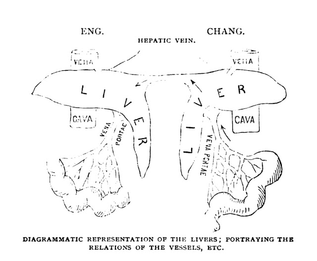
     The findings of the limited autopsy on the twins were reported during a meeting at the College of Physicians. The family permitted only the posterior aspect of the band to be opened, along with limited abdominal incisions. The major findings were that the twins shared liver tissue through the connecting canal, that their respective peritoneal membranes overlapped each other in the canal but remained separate, and that umbilical arteries terminated feebly at the entrance to the band (the umbilicus was considered shared in the center). Dye injected into the portal circulation on Chang’s side emerged on Eng’s side.

(From the article referenced below)

     During life it had become clear that the two arterial circulations and nervous systems were distinct. When Chang became intoxicated Eng did not, and if Eng ate asparagus only his urine smelled of it later. Similarly, if the center of the connecting band was pricked, they both felt pain, but not if pricked closer to their respective bodies.
  

     The autopsy satisfied the doctors’ curiosity but did not open any new doors. The bigger lesson, it seems, was that two conjoined individuals could tolerate each other for 63 years.


SOURCES:

Huang, Yunte. Inseparable: The Original Siamese Twins and their Rendezvous with American History. 2018; Liveright.

Report of the Autopsy of the Siamese Twins. (Reprinted from the Philadelphia Medical Times), 1874. Available on Hathi Trust.

Mall, F P. “On Some Points of Importance to Anatomists.” 1907; Science 25(630): 121-5.

Savitch, S L, et al. “Joseph Pancoast, MD (1805-1882): The Surgeon Who Brought Anatomy to Life.” 2020; The American Surgeon. (online only)

Rutgow, I. “American Surgical Biographies.” Surgical Clinics of North America, 1987; 67(6): 1153-80. 


Tuesday, March 16, 2021

                AMERICA’S FIRST GALLBLADDER SURGERY


     Mary E. Wiggins, a thirty-year-old seamstress, arrived in Indianapolis in 1867 to consult a surgeon about a severe pain in her right side. Four years earlier she had noted a lump near her right pelvic brim that grew in size and became tender. By the time of the consultation she had lost considerable weight and could not work. Eating and mere walking produced crippling pain. Her physician, Dr. Newcomer, thought she had a large ovarian cyst, as did other physicians that she consulted. Knowing that a number of these had been successfully removed, Newcomer brought her to see Dr. John S. Bobbs, a respected Indianapolis surgeon. 

Dr. John S. Bobbs (Ind Med J, Hathi Trust)

     Dr. Bobbs examined her carefully. He could only partially outline the mass and pelvic examination did not suggest any attachment to the uterus or its appendages. He told Mary that he doubted that this was an ovarian cyst. Desperate from pain, she begged for surgery, regardless of the diagnosis. Bobbs, uncertain, waited, but after a second examination revealed nothing new, he decided to operate. 

     He had Mary taken to a room on the third floor over a local drugstore, a room that he rented at times for surgery. Inside were a plain wooden table for operating, a bed, a chamber pot, and chairs. At the time there were no hospitals for private patients and no trained nurses available. Present at the surgery were Dr. Newcomer, five other physicians, and a medical student, the nephew of Bobbs’ wife. Chloroform anesthesia was given. 

     Dr. Bobbs made an incision from the umbilicus to the pubis. Because of numerous adhesions he had to push his gloveless fingers through to reach the mass. It was about five by two inches, had a thin, translucent wall, and appeared to be attached to the liver. He opened the lower end and clear, colorless fluid gushed out, ejecting several small concretions ranging in size up to that “of ordinary rifle bullets.” After removal of additional stones, Bobbs sutured the open end of the gallbladder to the abdominal wall to allow further drainage. Antisepsis was still unknown, but Bobbs was said to always wash his hands before surgery. 

Mary Wiggins, 38 yrs after surgery
(Ind Med J, Hathi Trust)

 An Englishwoman was hired to provide nursing care. Aside from a small stitch abscess, Mary Wiggins recovered uneventfully. Her portrait shown nearby was taken thirty-eight years after the surgery. 

     Dr. Bobbs was born in Green Village, PA, in 1809. He grew up speaking Pennsylvania Dutch, a German dialect, and learned English a little later. He was apprenticed to a Doctor Martin Luther, practiced a short time, and moved to Indiana in 1835. He later enrolled in a one-year course at Jefferson Medical College. Another pupil while at Jefferson was J. Marion Sims, later famous as a gynecological surgeon and important to this story. Bobbs returned to Indianapolis where, in 1854, he contracted cholera. His physician, as treatment, rubbed morphine on his tongue, gave frequent small feedings of crushed ice and champagne, and applied hot water bottles and massages to his cramping extremities. He survived.

     Dr. Bobbs was active in the establishment the Indiana State Medical Society and of the Indiana Medical College. He became professor of surgery at the College and later dean of the school. A medical colleague, while praising his surgical talent, added that “he was original and bold almost to recklessness.” He served with distinction in the Civil War, read widely, and served one term as a State Senator. He published a report of his innovative operation in 1869 and died in the following year.

     There appear to be no further cholecystotomies until 1878, ten years later. In that year his old schoolmate, J.Marion Sims, unaware

J Marion Sims (from Meine
Lebensgescgichte, 
Hathi Trust)

of Bobbs’ report in an obscure journal, performed a similar operation in Paris. The patient was a 45-year-old woman with pain and a palpable mass, complicated by jaundice and severe itching. Sims aspirated the tender mass, removing 32 ounces of dark fluid. This relieved the pain and itching, temporarily. When symptoms recurred, Sims opened the abdomen, employing the new Lister technique of carbolic acid washes and sprays. He removed 24 ounces of dark fluid and 60 gallstones from the gallbladder and sewed the edges of the empty sac to the abdominal wall. The patient did well for five days, then began to ooze blood from the wound, gums, and intestinal tract, dying of internal hemorrhage. (Possibly a clotting disorder?) At autopsy the wound was intact and the gallbladder was not distended. Sims concluded that the operation probably had a future as a remedy for painful gallbladder distension and possibly liver abscesses and hydatid cysts. He coined the word “cholecystotomy”, a mixture of Greek words for gall, bladder, and incision. 

     The following year, the English surgeon, Lawson Tait, reported another case of cholecystotomy to the Royal Medical and Chirurgical

   Lawson Tait  (from biography    
by McKay, Hathi Trust)

Society. This patient survived the surgery. In 1889 Tait reported on an extended experience with 55 cases, seven of whom died. Of interest, Tait, like Sims, was primarily a gynecological surgeon. 

     The first actual removal of the gallbladder was done by Carl Langenbuch, in Berlin. In 1882, after trial operations on animals, he removed the organ from a man whose chronic pain over years had reduced him to morphine addiction and a skeletal appearance. The day after surgery the man was reported to be pain-free and smoking a cigar. Modern gallbladder surgery was born.


SOURCES:

Brayton, A W. “John S. Bobbs of Indianapolis: The Father of Cholecystotomy.” 1905; Indiana Med J 24: 21-37.

Tinkere, M B. “The First Nephrectomy and the First Cholecystotomy, with a Sketch of the Lives of Doctors Erastus B. Wolcott and John S. Dobbs.” 1901; Johns Hopkins Hosp Bull 12: 247-51.

Bobbs, J S. “Case of Lithotomy of the Gall Bladder.” 1868; Trans Indiana State Med Soc 18: 68-73.

Sims, J M. “Cholecystotomy in Dropsy of the Gallbladder.” 1878; Brit Med J June 8, pp 811-15

Tait, L. “Case of Cholecystotomy Performed for Dropsy of the Gallbladder.” 1879; Proc Royal Med Chirurg Soc 9: 435-7.

McKay, W J S. Lawson Tait: His Life and Work. 1922; William Wood and Co.

Thorwald, Jurgen. The Triumph of Surgery. 1957; Pantheon Books.

Wednesday, February 17, 2021


Troops ready to land 
(Wikipedia)
WHAT HAPPENED TO THE TYPHUS                 VACCINE?


      America’s baptism of fire in WWII was “Operation Torch”, the code name for the invasion of North Africa. The troops faced numerous hazards. Among them was epidemic typhus, a louse-transmitted scourge in the local population, likely to infect troops that cannot easily bathe or change clothes. Acquired by rubbing the infected feces of body lice into skin during scratching, epidemic typhus had swept away millions during and after the previous World War. Fearing that another epidemic could ruin the upcoming invasion, planners reached for two weapons: insecticides and vaccination. 

     On the advice of the newly-formed U.S.A. Typhus Commission (USATC), directed by Brig. Gen. Leon Fox (chief military health officer for the North Atlantic) and Fred Soper (an epidemiologist instrumental in malaria and yellow fever control in South America), all invading troops carried two-ounce cans of MYL powder, a pyrethrin-based insecticide, and all received a new and as yet minimally-tested vaccine against typhus. 

     The journey to a vaccine began in 1909 with the discovery by Charles Nicolle, in Algeria, that body lice transmitted the unseen germ. The next year, studying typhus in Mexico, Howard Ricketts

Howard Ricketts (Wikipedia)

found the tiny organism in lice, a finding soon confirmed by others. Early in WWI, A Brazilian scientist working in Germany, Henrique da Rocha Lima, and a Czeck scientist, Stanislaus von Prowazek, both working in a camp holding Russian prisoners, many with typhus, visualized the tiny causative organisms inside the cells lining the louse intestine. Rocha Lima named the organisms Rickettsia prowazeki after Howard Ricketts and von Prowazek, both of
Stanislaus von Prowazek(Wikipedia)

whom died of typhus during their investigations (Rocha Lima also contracted the disease but survived). Attempts at culturing failed, however.

     Inability to grow the organisms hampered vaccine development. A Polish scientist, though, did produce a vaccine in the 1930s, used on a significant scale. The scientist, Rudolf Weigl, in Lvov, Poland, invented a technique to inject Rickettsia organisms into the rectums of sterile lice using a fine glass tube and operating under a binocular microscope. Lice were placed in small boxes strapped to the legs of humans for feeding, both before and again after anal injection, to

Rudolf Weigl (Wikipedia)

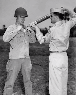
enhance growth of the Rickettsia. Weigl then delicately removed the infected intestines, ground them with phenol as a sterilizer, and used the material as vaccine fluid. Three doses over a month offered enough protection to modify or prevent illness or death. The vaccine was used in and around Poland and traveled to China, where it saved hundreds of missionaries. The Germans, after invading Lvov, realized the benefits of the vaccine for their troops. Ramping up production impelled Weigl to mechanize the louse injections, so that one operator could inject 50 lice at a time and about 1000 an hour. A skilled dissector 
Body louse feeding (CDC website)
could harvest 300 louse guts per hour and one person could feed about 25,000 lice a month.  During WWII Weigl, under a lenient German manager, also managed to smuggle 30,000 doses to ghettoes in Poland without discovery. A similar lab operated in Warsaw. Being a louse feeder or technician in the lab was a life-saving job for many where SS troops roamed the streets. 

Weigl vaccine, packaged (Wiki-Commons)

     After the war scientists attempted to grow the organism in animal tissues such as rodent lungs, with some success. French workers in North Africa developed such vaccines and tried another made from murine typhus organisms, a related Rickettsia that can cause mild human disease. More importantly, Herald R. Cox (after whom 

Herald Cox (Wikipedia)

Coxiella burnetii, the cause of Q fever, is named), of the U.S. Public Health Service, succeeded in growing R prowazeki in the yolk sac of chick embryos. Further steps, especially extraction by ether, led to a more potent and easier to make vaccine, eventually produced by Squibb and Lederle in the U.S. and Connaught Laboratories in Canada. Human trials undertaken in Bolivia and Spain gave indefinite results, but the U.S. military decided, to be safe, to vaccinate the troops invading North Africa. In spite of a severe epidemic in the area only a handful of troops came down with mild cases. Proper studies among subjects in Cairo proved that the “Cox vaccine”, as it was known, was effective in protecting against severe, though not all, disease. 

     The MYL powder eliminated lice from clothing easily but its potency died out quickly. The next year, though, DDT came into use. This seemingly magic chemical had been synthesized as early

Paul Müller (Wikipedia)

as 1874 but had no useful function. In 1939, Paul H. Müller, a chemist working at the Swiss chemical firm, J.R. Geigy, while looking for a good insecticide, discovered the superior properties of DDT. Compared to MYL it had a much longer duration of action and soon replaced it. It was in use in Africa by early 1943. For delousing, the military ordered the local population to remove their clothing. Moslem women refused, however. So, Army personnel employed spray guns to blow the chemical between the clothing and skin, with the sleeves and legs tied off. 
Spray gun for DDT (Wikipedia)

     Many millions of lives were saved with DDT. Paul Müller was awarded the Nobel Prize in Physiology or Medicine in 1948 despite the fact that he was neither a physician nor a physiologist.

     The Weigl vaccine, after the war, gave way to the easier “Cox vaccine” whose production continued until 1970. By then, the widespread use of DDT and the introduction of effective antibiotics had terminated typhus as a world problem. The former scourge of armies, prisons, ships, and famine presently exists only in isolated pockets. 


SOURCES:

Allen, Arthur. The Fantastic Laboratory of Dr. Weigl. 2014; W W Norton

Snyder, J C. “Typhus Fever in the Second World War”. 1947; California Medicine 66 (1): 3-10

Ecke, et al. “The Effect of Cox-Type Vaccine on Louse-Borne typhus Fever.” 1945; Am J Trop Med25: 447-62.

Lindenmann, Jean. “Typhus Vaccine Developments from the First to the Second World War.” 2002; Hist Phil Life Sci. 24: 467-485.


Friday, January 15, 2021


                 INTERNATIONAL MEDICAL CONGRESS OF 1876


     In 1876 the United States was 100 years old. To celebrate, Congress authorized a mammoth celebration, an international exhibition in Philadelphia, covering about 285 acres. The Exposition included a medical exhibit: a military hospital, fully built, with pathology specimens, books, instruments, etc. on display. In one room was a scale model of a medical railroad train, with beds and stretchers in some cars and an operating suite, kitchen, and baggage space in others. Such trains were used in the Civil War. Bright red paint on the smokestacks and other locations and red lights at night served as a signal not to attack it, and it never was.


Main building, Centennial, covers 21.5 acres (Wikipedia)

   The Philadelphia County Medical Society, not to be outdone, decided to hold an international medical congress concurrently with the Centennial. Doctors from Europe to Japan arrived, along with delegates from the AMA, state medical societies (delegates limited to the number of representatives in Congress), the three military services (Army, Navy, and Marines), the American Public Health Association, a mental hospital association, and the American Association for the Cure of Inebriates. It ran for six days in September 1876.

     The president of the Congress was Samuel D. Gross, the prominent Philadelphia surgeon, celebrated in a painting by Thomas Eakins. Joseph Lister chaired the section on surgery, where he

The Gross Clinic, by Thomas Eakins (Wikipedia)
It hung in the military hospital exhibit (paragraph 
one)

presented his new antiseptic approach to surgery. Alfred Stillé presided over the internal medicine section. He was professor of the theory and practice of medicine at the U. of Penn Medical School and had published an early American text of pathology but was lagging in his acceptance of germ theory.  Robert Barnes, of London, obstetric physician at St. George’s Hospital, presided over obstetrics. Various other specialties were represented, including “sanitary science” (public health, today) and mental illness. The program featured eighty-one papers.

     The Congress met at a time when new findings of bacteriology were upending long-held beliefs and practices. After Joseph Lister lectured on his antiseptic surgery technique, employing carbolic acid liberally as the key antiseptic, attendee comments made it clear that

Joseph Lister (Wikipedia and Wellcome Library)
those who had actually visited Lister in Scotland and seen his results were quite persuaded by the method, as were several more. Others were still doubters and professed to have good results with mere cleanliness. Samuel Gross, unconvinced by Lister, remained silent.     

     In the obstetrics section, puerperal fever was reviewed by William T Lusk, chief of obstetrics at Bellevue Medical College. He agreed on the presence of coccal bacteria in the tissues of puerperal fever victims but was unsure of the mechanism of sepsis. He discussed Semmelweiss’ successful trial of handwashing, adding that he had queried three attendings that did both autopsies and deliveries concurrently. Two had not encountered puerperal fever and one said his death record was “unusually low.” Lusk, conceding that bacteria played a role, concluded that puerperal fever did not have a single cause.

     In fact, there was no bacteriology laboratory in the United States at the time. Even in Europe the science was just beginning and confusion reigned over various bacterial species. Agar plates did not arrive until 1882, allowing more exact speciation. Petri dishes came a few years later.

     Dr. Thomas Satterthwaite, from New York, discussed the germ theory in relation to Sanitary Science (public health). He was equally confused, admitting some relation of certain bacteria to certain diseases, but noting that the air was full of bacteria, with no ill effects. Dr Woodworth, of the Marine Hospital Service, discussed quarantine. He referred to the “morbific causes of infectious diseases”, recommending overall hygiene, cleansing of ships and ports, as well as the presence of informants in foreign ports to report contagious diseases, an innovation later adopted.

     A few other items are of interest. Joseph Woodward, author of a medical and surgical history of the Civil War, noted an average mortality rate from disease (not wounds) among white volunteers in the Civil War of 55 per 1000 mean strength. Among blacks in the last two years of the War, the rate was 211 and 139.8 per 1000. The excess in blacks he attributed to their poor condition before enlistment. As fugitive slaves they were “literally naked and starving”, as he put it. 

     Mercury and potassium iodide remained the treatments for syphilis, with much discussion over dosage and regimens. Dr. George A. Ward, medical director of the Oroya Railroad in Peru, gave an interesting account of verrugas, a stage in what is now known as Bartonellosis, seen in railway workers in the Andes. The Railroad was a venture of Henry Meiggs, the builder of the huge gold-rush-era Meiggs Wharf in San Francisco (burned in the earthquake). He had fled to South America in 1854 to escape crushing debts, only to take up railroad building. 

     A paper on treatment of ulcers recommended taking milk, especially cow’s milk. For bleeding ulcers, better to swallow ice and apply it over the epigastrium. Other medical papers covered “typhomalarial fever” (still considered an entity), diphtheria and the history of American medicine and surgery. 

     Presentations on mental illness dealt with the histology of general paresis (syphilis of the brain) before the infectious cause was understood, the insanity defense in criminal cases, and institutions for care of the insane. Dr. Edward R. Squibb, founder of the Squibb company and an advocate of better drug purity, spoke on the hope of a “universal pharmacopeia”, to standardize medicines worldwide. He realized it was unrealistic at present.

     Except for that of Lister, there were no breakthrough papers at the Congress, and Lister’s presentation had a limited immediate effect on surgical practice. But the seeds of progress had been sown and soon sprouted in surgery's younger generation. 


SOURCES:

Ashhurst, J, ed. Transactions of the International Medical Congress, 1876

Rutgow, I. “Joseph Lister and His 1876 Tour of America”. 2013; Ann Surgery 257: 1181-7.

Maass, J. “When the New World Dazzled the Old” American Heritage, June 1976, pp 25-8.

Review of International Exhibition, 1876, Official Catalogue. 1876; Amer J Medical Sciences 72: 236-9.