Friday, November 13, 2020

 Orthopedic Surgery Enters the Modern Age on          a Chance Observation

Roy A. Meals, MD

     For thousands of years, bone setters and doctors could not accurately diagnosis broken bones or differentiate such injuries from joint dislocations and torn ligaments. That changed with a chance discovery 125 years ago this month. Subsequently, doctors began using the new discovery to discount their long-held assumptions and were able to accurately diagnose skeletal diseases. 

     In his darkened laboratory on November 8, 1895, a German mechanical engineer and physicist, Wilhelm Röntgen, electrified a vacuum tube and happened to observe a strange glow coming from a nearby card coated with a photosensitive chemical. He placed his hand between the vacuum tube and the card and an image of his hand appeared. Over the following weeks he ate and slept in his laboratory and studied this unknown ray, which he labeled “X,” the mathematical symbol for an unknown. 
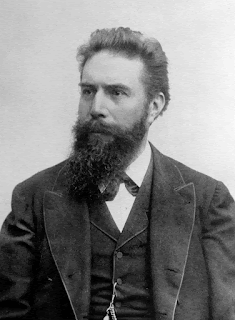
Wilhelm Röntgen (Wikipedia)

     Röntgen discovered that X-rays passed through books, no matter how thick, and that coins cast a shadow on the photosensitive board. Röntgen shared the secret with his wife, who allowed him to take a fifteen-minute exposure of her hand, the first orthopedic X-ray. When she saw the image of her hand skeleton, she exclaimed, “I have seen my death.” Far more broadly, she was witnessing the advent of diagnostic radiology and modern orthopedic surgery. 

     A week later, Röntgen presented his findings in a paper titled, “On a New Type of Rays.” This caught the immediate attention of physicists, who alerted the lay press. The discovery made the front-page headline news within a week of Röntgen’s public presentation. 

     At the time, vacuum tubes were well known and easy to make. After Röntgen’s discovery and announcement, many investigators contributed to the understanding and practical applications of X-rays. Interest was intense and advances were rapid. Less than three months after Röntgen’s public announcement, an enterprising electrical contractor and avid photographer opened a laboratory that offered diagnostic services. 

X-ray of Bertha's hand (Wikipedia)

     Röntgen received the Nobel Prize in 1901, the first one ever awarded for physics. Röntgen not only gave the reward money to his university, he also refused to take out patents on his discovery to allow for wide-spread application. 

     I suppose when X-rays were in their infancy, patients were asking, “Now that you have finished obtaining a thorough medical history, performing a careful physical examination, and telling me that you know with assurance what is wrong, aren’t you going to order an X-ray, Doctor?” This question implied a lack of trust in the doctor’s diagnosis unless he threw in a high-tech, oh-so-modern X-ray evaluation. Gradually, doctors and patients came to understand when an X-ray study could help make the diagnosis or plan treatment and when one would be superfluous. For instance, today it is intuitive that a sore tooth most likely deserves an X-ray while a sore throat does not. In general, X-rays reveal calcium-rich structures—those containing enough calcium to cast a shadow in the X-ray beam. Examples are bones, teeth, hardened arteries, and kidney stones.

     Doctors have learned to order X-rays with some caution because radiation damages living tissues and their DNA. That fact required discovery, and the harmful effects of early X-ray examinations were slow to reveal themselves. Since X-rays could not be seen or felt, investigators had no reason to consider them harmful. Both Nicola Tesla and Thomas Edison experimented with X-rays, and both observed that their eyes became irritated; but neither drew a connection between the radiation and their symptoms. 

     For convenience, dentists originally held the film inside the patient’s mouth with their fingers when shooting dental X-rays. Decades later the skin on their hands dried, cracked, and became cancerous. Nowadays the radiology tech steps behind a lead shield before shooting the film, and there are generally accepted standards for how much radiation a person can receive on an annual and lifetime basis without incurring undue risk. 

     Despite our best efforts, we cannot avoid radiation exposure entirely. Some comes naturally from the sun and some from the ground. We get more during a plane flight because the thinner air at high altitude blocks less of the sun’s radiation. This fact poses a major, unsolved problem for interplanetary travel because of the absence of Earth’s radiation-shielding atmosphere and because of the impracticality of armoring spaceships with lead. 

     Most would agree, however, that the potential benefits of a timely chest X-ray or mammogram far outweigh the risks.  Even an occasional and judiciously planned CT scan may help maintain or restore one’s health; but we should avoid advice such as, “I don’t have a clue about what’s wrong, so let’s get a CT scan.” A second opinion is safer. Remember, it took decades for the damaged DNA in dentists to turn into skin cancers. Similarly, we should avoid being our own doctor and proclaiming, “I would just feel better, Doctor, if you ordered a CT scan.”

     Röntgen discovered his new type of ray a few decades after the introduction of general anesthesia and the acceptance of aseptic surgical techniques. These developments, along with the invention of stainless steel, ushered into the modern era orthopedic surgery and the practicality of operative fixation of fractures. Looking ahead 125 years, fractures will still exist. Arthritis and osteoporosis may be fully preventable. Bone imaging techniques will be even more sophisticated than those in current use. X-ray imaging may be obsolete, replaced completely by magnetic resonance imaging, ultrasound, or some yet-to-be-discovered alternative. Nevertheless, Röntgen’s discovery and its enduring 125-year legacy deserves recognition and respect. 

Bradley, William. “History of Medical Imaging.” Proceedings of the American Philosophical Society 152, no. 3 (2008): 349–61​.

“Radiation Risk from Medical Imaging.” Accessed November 6, 2020. https://www.health.harvard.edu/cancer/radiation-risk-from-medical-imaging

Röntgen, William. “Ueber eine neue Art von Strahlen. (On a New Kind of Rays.)” In Classics of Orthopaedics. Edited by Edgar Bick, 278–84​. Philadelphia: Lippincott, 1976.

Dr. Meals blogs at www.aboutbone.com and has recently published Bones, Inside and Out (WW Norton, 2020) 

Wednesday, October 14, 2020


 Presidential Disability: 

The Case of Grover Cleveland and the

Twenty-fifth Amendment


     On June 30, 1893, President Grover Cleveland called for a special session of Congress to meet on August 8. The economy was slumping badly and he urgently wanted Congress to repeal the Sherman Silver Purchase Act. But why wait until August? Because he had scheduled a leisurely cruise up the Hudson River to his summer retreat at Buzzards Bay, he said.

     The truth was different. In May, Cleveland had noticed a roughening in his mouth. An examination by Dr. Robert O’Reilly, his personal physician, revealed a quarter-sized, ulcerating lesion on the roof of his mouth. Dr. Joseph Bryant, from Bellevue Hospital

Dr. Joseph Bryant (Nat. Library of 
Medicine)

Medical College, took a biopsy and sent it to William Welch at Johns Hopkins. Welch considered the tissue malignant, and Bryant urged immediate surgery. Cleveland felt, considering the economic crisis, that news of an operation for cancer would be destabilizing. Accordingly, he chose to have the tumor removed secretly aboard a yacht, the Oneida, loaned by a wealthy friend. 

     On the morning of July 1, 1893, the Oneida left the New York pier. On board, beside the President, were the surgeon, Dr. Bryant, who had authored a textbook on surgery and had reported the world’s largest series of maxillary (upper jaw, roof of mouth) resections, John Erdmann, a surgeon who worked with Bryant, Dr. William Keen, a well-known neurosurgeon from Jefferson Medical College, Edward Janeway, a prominent

Dr. Edward Janeway (Wikipedia)

internist from Bellevue, Ferdinand Hasbrouck, a dentist and anesthesiologist, and Dr. O’Reilly, the President’s personal physician who was to administer ether.

     The surgery began at 12:50 PM. Anesthesia was a worry, as the President was fifty-six years old, was obese, and had a short, thick neck. Initially, under nitrous oxide and locally injected cocaine anesthesia, Hasbrouck extracted two teeth and Bryant made an initial incision over the hard palate. Then, as Reilly administered ether, almost the entire upper jaw was removed along with some soft palate. A large gelatinous mass in the maxillary sinus was diagnosed by Dr. Welch as carcinoma. Gauzes were applied and the patient awakened. The surgery lasted just over one hour, with about six ounces of blood lost. Just over two weeks later Cleveland returned to the Oneida for a resection of margins thought to harbor residual tumor.

     The President recovered at Gray Gables, his home in Buzzards Bay. His speech was muffled and indistinct and his left cheek hollow. The team requested Dr. Kasson Church Gibson, a New York dentist, to make a prosthesis. Gibson had studied with Dr. Norman Kingsley, known as the “father” of orthodontics. Kingsley was the first to use a vulcanized rubber obturator (an object to fill a gap) in a case of cleft palate. He founded the New York College of Dentistry and wrote the first text on orthodontics. His apprentice, Gibson, after dentistry school, opened a practice in New York and was on the faculty of the University of Maryland School of Dentistry. He had collaborated with Bryant on previous cases. Gibson produced an obturator so perfect that the President’s face and speech appeared normal, even to friends, and he had no difficulty eating.


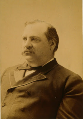
Pres. Cleveland, 1904 (Smithsonian
Portrait Gallery)
Pres. Cleveland, 1888 (Library of
Congress)


     In 1980, pathologists from the University of Pennsylvania restudied the tumor, preserved in the Mutter Museum in Philadelphia. They decided that the tumor was a verrucous carcinoma, a slow-growing tumor not prone to metastasis. Cleveland was probably cured at the initial surgery. 

     To explain the President’s temporary weakness and weight loss, Cleveland’s staff gave out reports that he suffered from rheumatism and bad teeth. One reporter published a true account, obtained from an inadvertent slip by Hasbrouck, but the coverup was otherwise so complete that the report never gained credence and the full story did not emerge until after Cleveland’s death.


     Coverups of presidential illness have been fairly common. Concealing the illnesses of Wilson, Roosevelt, Eisenhower, and Kennedy are examples. Most recently, when President Trump was hospitalized, the issue of a temporary incapacitation arose. A mechanism to deal with incapacity of the president, temporary or otherwise, is contained in the 25th amendment to the Constitution, written well after Cleveland’s time.

     The idea of a plan in case of temporary presidential disability originally came from President Eisenhower. Eventually, Lyndon Johnson formally asked Congress to address the issue with a constitutional amendment. The amendment from Congress was ratified in February 1967. But does it address a central issue?

     A radiologist at Stanford University, Herbert Abrams, has pointed out that Congress never solicited medical opinion (from the AMA, for example) when drafting the amendment. In addition, the amendment says nothing about obtaining medical consultation in cases where a president is unable to perform his duties but will not or cannot say so. It reads only that “the Vice President and a majority of the principal officers of the executive departments or of such other body as Congress may by law provide” transmit to Congress their written declaration of the President’s incapacity. Abrams felt that though medical advice would almost certainly be sought, the mechanism should be spelled out. He suggested a six-man panel of physicians consisting of two internists, two neurologists, a psychiatrist, and a surgeon to provide an objective medical opinion if needed. Each would serve a six-year term, with a rotational replacement schedule, the appointments to be made by a complicated process to ensure impartiality.

     Perhaps renewed discussions in Congress will lead to clarification of this issue. Meanwhile, since staffs are generally loyal to their presidents and a cabinet majority is needed to invoke the 25th amendment, some degree of coverup may well be a feature of future presidencies.


SOURCES:

Ferrell, Robert. Ill-Advised: Presidential Health and Public Trust. 1992; Univ of Missouri Press.

Keen, W W. The Surgical Operations on President Cleveland in 1893. 1917; George Jacobs Co.

Seelig, M G. “Cancer and Politics: The Operation on Grover Cleveland” 1947; Surg Gyn Obst 85: 373-6.

Murray M, Pappas T, Powers D. “Maxillary Prosthetics, Speech Impairment, and Presidential Politics: How Grover Cleveland Was Able to Speak Normally after His “Secret” Operation.” 2020; Surg Journal 6: e1-e6.

Abrams H. “Can the Twenty-fifth Amendment Deal with a Disabled President? Preventing Future White House Coverups.” 1999; Presidential Studies Quarterly 29 (1): 115-33.      


Tuesday, September 15, 2020

                                        SAMUEL POZZI

FATHER OF FRENCH GYNECOLOGY


     In the summer of 1881 an up-and-coming artist, John Singer Sargent, was introduced in Paris to a rising French physician, Samuel Pozzi. The artist agreed to do a portrait of Pozzi,

Dr. Pozzi at Home by J S Sargent
(Wikipedia)
a strikingly handsome man. When Sargent visited Pozzi’s sumptuous home on the Place Vendome he found Pozzi in a bright red dressing gown and decided on the spot to portray him in this garment. The result was one of Sargent’s most arresting paintings.

     Who was Samuel Pozzi? Born in 1846 to a physician father in Bergerac (of Cyrano fame), he completed his medical education at the University of Paris. During the student years he lived in the Latin Quarter, where he met the young actress, Sara Bernhardt. The two were almost certainly lovers and remained close friends throughout life. 

     In 1870 the Franco-Prussian War broke 

Samuel Pozzi (Wikipedia)
 out. Pozzi, then an intern at the Necker   Hospital, enlisted and treated the wounded at  Sedan. After the war he completed medical school, writing a thesis on fistulas of the upper recto-pelvic space. His aggregation thesis, for a faculty position, was on fibroid tumors of the uterus. Pozzi then worked at the Pitié Hospital, whose director was Paul Broca, a noted anatomist, surgeon, and anthropologist, who had discovered the speech area of the brain, “Broca’s area”. At Broca’s encouragement, Pozzi and a colleague translated into French Darwin’s latest work, The Expression of the Emotions in Man and Animals

    To enhance his income Pozzi took locum tenens positions at several hospitals and  gradually built a private practice. In 1876, at a meeting of the British Medical Association, he visited Joseph Lister in Edinburgh. He learned his techniques and on return introduced them into his work at the Pitié, supported by Broca. He was one of the early users of antiseptic techniques in France. 

     In 1883 he was appointed chief of surgery at the Lourcine-Pascal Hospital (later called the Broca Hospital). Pozzi had been interested in medical and surgical problems of women since medical school, where he had been impressed at how neglected their problems were. Particularly distressing were fistulas, leaking urine or stool, seen after difficult childbirths. Pozzi converted a disused shack on the grounds into a specialized ward for gynecological patients, considered to be the first such unit in France. Pozzi also installed an operating room in the main hospital, something it was lacking, and acquired possibly

Pozzi operating at the Broca Hospital (Standing to right of patient) 
(Wellcome Library and Wikicommons)

the first autoclave in France. Believing in the healing power of art, Pozzi had murals painted on the ward walls and a ceiling painted by the artist Georges Clairin, entitled Health Restored to the Sick. Sarah Bernhardt was the model for “Health”. A new surgical assistant, Robert Proust, brother of Marcel the writer, joined him. Proust later
Robert Proust (Wikipedia)

made contributions to gynecological and urological surgery.

     Pozzi applied to the University of Paris to teach a course in gynecology but was refused, so he taught the first such course in France independently and free of charge. It was popular. Meanwhile he worked to accumulate material for a much-needed textbook of gynecology. He finally retreated to his mother-in-law’s house in Montpellier for six months of isolated labor to produce it. 

     The text, the first in France, published in 1890 in two volumes and comprising over 1200 pages with almost 500 illustrations, was immensely successful and was followed by a new edition two years later. New surgical instruments and techniques that Pozzi had introduced were important contributions. The work was translated

From Pozzi's Textbook, 1891
Translation (Hathi Trust)


into five languages, including more than one English edition and new editions followed. The text established the specialty of gynecology in France and enhanced it worldwide. Amazingly, it wasn’t until 1901 that a department of gynecology was established at the University of Paris, with Pozzi appointed to chair it. 

     In contrast, in the U.S. the American Gynecological Society was formed as early as 1876. In that year Ulysses Grant was president, Custer was killed at Little Big Horn, and Colorado was admitted as the 38th state. 

     Pozzi’s practice expanded, though he never abandoned general surgery, and he served as personal physician to numerous patients, including celebrities. He removed a large ovarian cyst from his friend Sarah Bernhardt but deferred to a colleague when she needed a leg amputation later in life. He continued to mingle with artists and writers and traveled frequently, accumulating a significant art collection. The American Gynecological Society honored Dr. Pozzi in 1906, the one hundredth anniversary of Ephraim McDowell’s amazing kitchen-table ovariotomy, by inviting him to address the Society on the history of ovariotomy in France. 

     Pozzi was deeply involved in the Dreyfus scandal, acting as a vocal advocate for the wrongly accused officer. He was also elected as a Senator for his home area. When WWI came, Pozzi enlisted at age 68, operating again on wounded soldiers and writing papers on abdominal wounds.

     Pozzi’s end came suddenly in 1918 when a deranged patient in his office shot him four times in the abdomen. Rushed to emergency surgery, Pozzi insisted on remaining conscious, ordering the surgeons to use only local anesthesia, morphine, and atropine. Prime Minister Clemenceau, an old medical school friend, attended the surgery. Despite repairing the bowel perforations, Pozzi died from a sudden hemorrhage. 

     The death of Samuel Pozzi, the creator of gynecology in France, was mourned by thousands, both for his work as a physician and for his support of, and friendship to, the artistic community of France. The famous portrait by Sargent is now on display in the Hammer Museum in Los Angeles.


SOURCES:

De Costa, C and Miller, F. The Diva and Doctor GodLetters from Sarah Bernhardt to Doctor Samuel Pozzi. 2010, ExLibris Corp.


Barnes, J. The Man in the Red Coat. 2020, Knopf.


Pozzi, S. Treatise on Gynecology, Medical and Surgical. Trans from French under supervision of Brooks H. Wells. 1891; Wm Wood & Co.


Speck, R. “Robert Proust – an eminent doctor in the shadow of his famous brother Marcel.” 2001; World J Urol 19: 285-91.


Ricci, J. The Development of Gynecological Surgery and Instruments. 1949; The Blackiston Co.


Pozzi, S. “The Evolution of Ovariotomy in France.” 1909; Surg, Gyn, Obs 9: 417-26.


Taylor, E S. History of the American Gynecological Society 1876-1981 and American Association of Obstetricians and Gynecologists 1888-1981. 1985; C.V. Mosby Co. (available online at: https://www.agosonline.org/public/PDF/Taylor_AGOS_history.pdf )


Transactions of the American Gynecological Society.  1876; H O Houghton & Co. vol 1.


Monday, August 17, 2020

                  THE BIRTH OF THE CALIFORNIA STATE

                               HEALTH DEPARTMENT 

      In epidemic times, such as these, we look to local or national health departments for information and guidance. But that wasn’t always the case. In earlier times health departments were ad hoc organizations formed on the spot to deal with epidemics as they arose, only to die as fears abated. Such was the case in San Francisco when cholera swept through the city in early gold rush days. In 1862, however, when smallpox broke out in Sacramento, the city formed a permanent health board that lasted to this day, and six years later the State of California followed suit with a permanent State Health Board. Driving the formation of both health boards was a practicing Sacramento physician, Thomas M. Logan.

     Born in Charleston, S.C., Logan graduated from the Medical College of South Carolina in 1828. He practiced locally for a short time, then travelled to Europe for further study, arriving in Paris at the time of

Thomas Logan (Cal West Med 1945)
the 1832 cholera epidemic, a valuable experience. After returning to America he opened a practice in New Orleans, which he abandoned in 1849 for gold in California. His voyage on a sailing vessel lasted nine months, four of them spent battling windstorms just to round Cape Horn, bringing him to San Francisco in January 1850. After a brief time at the mines, Logan opened a practice in Sacramento, then a town of about 6,500 people settled in a line of tents, primitive houses, and small businesses along the river. Cholera and scurvy were common, facilities primitive, and mortality high. 


Gold Rush Sacramento (Wikipedia)

     In 1858, the civic-minded Logan and Dr. Elias S. Cooper of San Francisco, founder of the first medical school in California, collaborated to form the California State Medical Society, the

        Elias S Cooper (Nat. Library of Medicine)    
   forerunner of the California Medical Association. The Society dissolved in 1860 over internal disputes and Cooper died in 1862, but Logan, with colleagues in Sacramento, resurrected the State Medical Society in 1870, to which he was unanimously elected president.

     That same year, prodded by a frightening smallpox outbreak in 1868-9, Logan induced the State Legislature to create a State Health Board, the forerunner of today's Dept. of Public Health. It was the second  permanent state health board in the nation and was modeled on the first one, created in Massachusetts only a year before.  A Health Board and quarantine service for San Francisco were created in the same year.

     The State Board’s task was to gather statistics of births, deaths, diseases, and the like. Weather, humidity, and geologic conditions were to be recorded, health conditions in public institutions, such as schools, prisons, and almshouses, were to be monitored, and the effects of intoxicating liquors on the personal and working lives of citizens (a serious problem at the time) evaluated, all to be summarized in a Biennial Report. This basic information was heretofore rudimentary and incomplete throughout the state. The Board was advisory and had no enforcement powers. It consisted of seven physicians, two from Sacramento and the others from diverse areas of the state, who would serve four-year terms. Logan was the Permanent Secretary, the only paid position, and Henry Gibbons of San Francisco was the president. Reporting forms were sent out to physicians and hospital personnel asking them to tabulate diagnoses and special medical problems. Interestingly, the Independent Order of Odd Fellows agreed to provide a monthly report of illness and death among its California members, because the IOOF contained “the most intelligent, sober, and industrious portion of our fellow citizens.”

Wyatt Earp's membership card to IOOF (Wikipedia)

     The Board’s first Biennial Report included reports on water supplies and purity, conditions in county hospitals, criminal abortion, the gathering of vital statistics, the unhealthy conditions in San Francisco’s Chinatown, and the “medical topography” of the state. The latter topic was of particular interest to Logan, who had personally measured temperature, humidity, and barometer readings since his arrival in California. The San Francisco County Hospital was deemed a “disgrace” and the new suburban Sacramento

First report of State Board of Health 
(Google Books)
Hospital showed an unexpected increase in mortality in spite of cleaner suburban air. Water was examined microscopically and tested chemically, but not cultured.

     A Dr. Briggs in Santa Barbara lauded the city’s restorative climate and included a comment that “about one and a half miles from the shore is an immense spring of petroleum, the product of which continually rises to the surface of the water and floats upon it over an area of many miles.” He thought that the ocean wind blew in a petroleum-derived “disinfecting agent” that accounted for the rarity of epidemic disease in Santa Barbara. 

     The Biennial Report reflected the confused state of medical thought at the time. Germs were occasionally mentioned, but miasmas and “malaria” (in the sense of bad air) still dominated thought. Infectious diseases were the main killers in California, “consumption” claiming the most victims, partly because the perceived healthy climate of the State attracted numerous tubercular patients. 

     Logan was a member of the AMA almost from the beginning, served as its delegate to the International Medical Congress in Paris in 1867, and went on to be President of the AMA in 1872. Through the AMA he pushed for a national public health bureau but did not live to see it. He continued as Permanent Secretary of the California Board of Health until his death in 1876. 

     Logan made many contributions to medicine, but his drive for “state medicine” in California and the nation ranks highest. 


SOURCES:

Harris, H. California’s Medical Story. 1932, Grabhorn Press


Jones, J R. Memories, Men, and Medicine: A History of Medicine in Sacramento, California. 1950, Sacramento Society for Medical Improvement.


Dickie, W M. “National Department of Health Proposed in 1871: by Thomas M Logan, MD, of California. 1940; Calif West Med 52: 6-9.


Saunders, J B. “Geography and Geopolitics in California Medicine”. 1967; Bull Hist Med 41: 293-324.


Biennial Report Calif State Board Health 1870-71. Sacramento State Printer.


Logan, T M. “Report on Topography, Meteorology, Endemics, and Epidemics”. 1858; Trans Med Soc State of Calif. 3: 31-80.


Jones, G P. “Thomas M. Logan, MD, Organizer of California State Board of Health and Co-Founder of the California Medical Association”. 1945; Cal West Med 63: 6-10.

Thursday, July 16, 2020


GUY’S HOSPITAL AND MODERN MEDICINE


          In the years after the Napoleonic wars English medical graduates, forgetting their antipathy toward the French, rushed to Paris to study. There, they learned medicine at the bedside and followed their cases to autopsy, already a standard procedure to resolve clinical problems. Patients were examined with palpation, percussion, and auscultation, using the newly invented stethoscope. Old disease classifications based on symptoms were giving way to new terminology based on visible pathology.
Entrance to Guy's Hospital, 1820 (Wikipedia)

      Guy’s Hospital Medical School, in 1825, was considered the best in London. This was partly due to the influence of Sir Astley Cooper, a talented surgeon who taught anatomy and surgery, subjects that focused clinical teaching on local pathology. Guy’s was also blessed with three physicians who together, reflecting the French influence, catapulted the medical service into prominence: Thomas Hodgkin, Thomas Addison, and Richard Bright.

     Hodgkin, a Quaker, having studied in Edinburgh and Paris, arrived at Guy’s in 1825 as a clinical clerk. Focused primarily on pathology, he soon assumed charge of autopsies and directed
Thomas Hodgkin (Wellcome Library)
the anatomical museum. He introduced the stethoscope to Guy’s and he pushed for bedside teaching, not yet in vogue in London. His pathology studies culminated in a famous paper of 1832 describing seven cases of enlarged spleen and lymph nodes (an original illustration of one case, borrowed from the pathologist Robert Carswell, can be seen in: Arch Int Med 121: 288-90, 1968)). Only three or four of his cases would be labeled Hodgkins disease  today. He resigned from the staff at Guy’s in 1837 after a tense relationship with the hospital treasurer had cost him a promotion. He spent much subsequent time as a social reformer.

     Thomas Addison, another Edinburgh graduate, after working at the Carey Street Public Dispensary under Thomas Bateman (one of the fathers of dermatology), was appointed assistant
Thomas Addison (Wikipedia)
physician at Guy’s in 1824. At Guy’s he excelled as a lecturer, published on pneumonia, phthisis, fatty liver, and other subjects, and, having learned skin diseases from Bateman, started a department of dermatology. Of great importance was the publication, co-authored with Richard Bright, of volume one of a text entitled Elements of the Practice of Medicine, a book that the students devoured. Addison, who wrote most of it, described the pathology, including microscopic findings, of pneumonia and covered the clinical picture and pathology of appendicitis, a clinical entity not fully appreciated at the time. In 1855 he published a
n
Patient with Addison's disease. Note copper skin (Wellcome Library)

book on the effects of disease of the adrenals, describing the full clinical picture of “Addison’s Disease”, a name actually coined by Armand Trousseau. In it he included some cases we now call pernicious anemia.

     Richard Bright came from a wealthy family, studied medicine at Edinburgh, and worked at the London Lock Hospital (a
Richard Bright (Wikipedia)
hospital for venereal disease), then at two other public hospitals under Thomas Bateman, from whom he also learned dermatology. He was appointed to the staff at Guy’s Hospital in 1820, preceding Addison’s appointment by four years and Hodgkin’s by five.

     Bright’s first important publication was in 1827, entitled Reports of Medical Cases Selected with a view of Illustrating the Symptoms and Cure of Diseases by a Reference to Morbid Anatomy. Inside were numerous case histories with autopsy findings, complete with numerous color illustrations. The latter included depictions of chronically diseased kidneys from patients who would thereafter be described as suffering from “Bright’s Disease”, or “Morbus Brightii” in those days.
Chronic kidney disease, mezzotint. From Bright's "Reports" (Internet Archives)

     The book was stunning. Nothing comparable had appeared in British medical literature since Matthew Baillie’s text on morbid anatomy of 1793 (that was still in print). Baillie’s book, though considered a landmark in pathology, contained no illustrations and no clinical histories, just descriptions of pathology. (In 1799 a volume with etchings to supplement the text was issued.) Bright’s manuscript included the clinical picture with the pathological findings. More striking were the color illustrations, for which Bright hired a father-and-son pair
of artists, one a mezzotint engraver. Mezzotinting was an expensive procedure but provided beautiful results. Bright, who was wealthy, presumably paid for the art himself. Overall, the two volumes contain 40 plates, 34 of which are mezzotints. The costly books were out of reach of medical students, and only 114 copies were sold in the first three years.
Ileal ulcers in typhoid fever, from Bright's "Reports" (Internet Archives)


     Bright also found that the urine of his renal patients contained albumin, which he tested for by holding a teaspoon of urine over a candle flame. Coagulation signified the presence of albumin. Others found urea in the blood of his patients, supporting the findings of Prevost and Dumas who had demonstrated urea in the blood of animals whose kidneys had been removed (1823), advancing the idea that a function of kidneys was to remove urea from blood.

     In time special beds were set aside for further study of patients with renal disease, complete with a small laboratory. It is thought to be the world’s first clinical research unit.

     Bright resigned from the staff of Guy’s Hospital in 1844 and entered private practice. He received many honors and was appointed Physician Extraordinary to Her Majesty Queen Victoria. His patients included John Snow (discoverer of the source of cholera) and Alfred Lord Tennyson. He died in 1858 after periods of angina and breathlessness.

     These brief and incomplete sketches only hint at the remarkable trio that was instrumental in introducing the new clinic-pathological approach to medicine in England. The introduction of chemistry into the study of renal disease and the use of microscopy in pathology were other contributions of this trio that have benefited us to this day.


SOURCES:

Rosenfeld, L. Thomas Hodgkin: Morbid Anatomist, Social Activist. 1993. Madison Books

MacKenzie, J C. “Dr. Richard Bright: A man of many parts - His bicentenary year. 1989; Brit Medico-Chirurg J 104: 63-67.

Peitzman, S J. “Bright’s Disease and Bright’s Generation: Toward exact medicine at Guy’s Hospital” 1981; Bull Hist Med 55: 307-21.

Keith, N M and Keys, T E. “Contributions of Richard Bright and His Associates to Renal Disease”. 1954; Arch Int Med 94: 5-21.

Pearce, J M S. “Thomas Addison (1793-1860)”. 2004; J Roy Soc Med 97: 297-300.

Dale, H. “Thomas Addison: Pioneer of Endocrinology”. 1949; Brit Med J Aug 13: 347-52.

Images of Hodgkin’s stethoscope and a model of Addison’s Disease are available at The Gordon Museum at King’s College London : https://www.kcl.ac.uk/gordon/collection/specimens - HodgkinsDiseaseSpecimen

Hodgkin, T. “On Some Morbid Appearances of the Absorbent Glands and Spleen.“ 1832; Medico-Chirurgical Transactions 17: 68-114.

Bright, R. Reports of Medical Cases Selected with a View of Illustrating the Symptoms and Cure of Diseases by a Reference to Morbid Anatomy. 1827; Longman, London.
Baillie, M. The Morbid anatomy of Some of the Most Important Parts of the Human Body. 1812 edition. Nicol and Johnson, London

Monday, June 15, 2020


America’s First Bronchoscopist


     Imagine a young girl, perhaps 7 or 8 years old, pale, weak, and emaciated. Her distraught mother explains that weeks earlier, her daughter had swallowed lye, thinking it was sugar. The resultant scars in her esophagus, contracting inexorably, now placed her near death from starvation or dehydration. During much of the 19th century most homes kept lye for making soap, and the jars did not carry danger labels. Other home accidents included aspirated or swallowed bones, safety pins, or coins, that might obstruct or perforate the esophagus or bronchial tree. Short of chest surgery, risky at the time, there was little relief available.

     To the rescue came the inventive Dr. Chevalier Jackson. Born in 1863, Jackson grew up on a poor farm near Pittsburgh.
Chevalier Jackson (Wellcome Library)
Valuing education, he worked his way through Western Pennsylvania University (now Univ of Pittsburgh) and entered Jefferson Medical College. He took an interest in laryngology, influenced by professors at the recently founded nose and throat clinic. In the library of one professor, Jackson read through the works of Sir Morrell MacKenzie, the father of laryngology in England. Determined to study with MacKenzie, he booked the cheapest steerage passage to England, requiring him to bring his own mattress for the assigned double-decker bunk. He stayed with MacKenzie only briefly, however, disappointed in the design of his esophagoscope.

     On return, in 1886, he opened an office in Pittsburgh, a coal town that was so sooty that the lights were often kept on throughout the day. Jackson’s new specialty of laryngology, fortunately, was publicized by news bulletins about the crown prince of Germany, the son-in-law of Queen Victoria, who had cancer of the larynx and on whom MacKenzie had consulted (see blog of 2/11/17).

     Jackson was busy very soon. Enlarged, chronically infected tonsils interfered with sleeping and breathing in those days, and he removed them from many poor public-school children, rarely receiving any payment. He also was adept at passing a tube through the larynx or doing tracheotomies in children with diphtheria (where an anesthetic was contraindicated). His inventive genius, however, found its greatest application while dealing with foreign bodies and lye burns of the esophagus. Within four years of arrival he had devised a new esophagoscope, with which he removed a “tooth plate” from the esophagus of an adult and a coin from that of a child. He reported the cases to the local medical society, only to learn of tragic outcomes in similar instances treated by his untrained colleagues. He refused henceforth to allow the instruments to be used without proper instruction in anatomy and practice on anesthetized dogs.

     When Jackson found he could dilate constricted esophagi with his instruments, unexpected numbers of emaciated children crowded his office. The numbers were so large that he initiated a crusade to have labels placed on containers with poisonous substances. Industrial resistance to labeling was fierce, however, and no such law was passed until 1927 – the Federal Caustic Poison Law.

     Meanwhile in Austria, in 1897, Dr. Gustav Killian, the “father of bronchoscopy”, made history by removing a piece of bone
Gustav Killian (Wikipedia)
from the bronchus of a farmer, using a rigid tube. Killian’s reports stimulated Jackson to devise his own “bronchoscope”, and, in so doing, he introduced bronchoscopy to America. Lighting in the distal end was adapted from cystoscopy techniques. Jackson’s rigid scope was used almost exclusively for foreign body retrieval and biopsies.

     Jackson continued to revise and perfect new models of his scopes. He was rigorous about requiring training for their use, and many of those he
Swallowed trinket embedded in a candy, X-ray by C. Jackson (Wikipedia Commons)
trained went on to teaching positions elsewhere. As his reputation spread, he was asked to demonstrate at conferences in major cities, including Paris. He bypassed London, however; England’s severe antivivisection laws prohibited endoscopy on dogs, even anesthetized ones.

     In 1916, he accepted the Chair of Laryngology at Jefferson Medical College in Philadelphia, and eventually held faculty positions simultaneously at five medical schools in Philadelphia. This broke exclusivity rules at most of the institutions, but regulations were bent to keep him aboard. His one stumbling block was tuberculosis, each of three episodes putting him at bedrest for months at a time. He used the idle time to
Title page of Jackson's text (Hathi Trust)
write a popular text on endoscopy, a revised edition for use as a manual, other texts, and many articles, 238 of which were single-authored.
He was an editor of the Archives of Otolaryngology, wrote an
autobiography, and was a founder of the American College of Surgeons. In his lifetime he extracted from patients over 2000 foreign bodies, all in the Mütter Museum in Philadelphia.

     Beneath this busy and inventive exterior lay a humane and charitable soul. Chevalier Jackson maintained that ninety percent of his work with patients was gratis. He never patented any of his instruments, considering it immoral to profit from medical inventions. He was a vegetarian and attributed his unusually steady hand in bronchoscopy to his abstinence from alcohol and tobacco. He was an accomplished artist and woodworker. He died at his farm, Sunrise Mill, near Philadelphia, at the age of 93.
Sunrise Mill, now a historic site (Wikipedia)


SOURCES:

Jackson, C. The Life of Chevalier Jackson: An Autobiography. 1938; Macmillan, 1938.

Marsh, B R. “Historic development of bronchoesophagology”. Otolaryngology-Head and Neck Surgery, 1996; 114: 689-716.

Boyd, A D. “Chevalier Jackson: The father of American bronchoesophagoscopy”. 1994; Ann Thoracic Surg 57: 502-5.

Coates, G M. “Chevalier Jackson” (obit.) 1959; AMA Arch Otolaryng 69(3): 372-4.