Monday, August 17, 2020

                  THE BIRTH OF THE CALIFORNIA STATE

                               HEALTH DEPARTMENT 

      In epidemic times, such as these, we look to local or national health departments for information and guidance. But that wasn’t always the case. In earlier times health departments were ad hoc organizations formed on the spot to deal with epidemics as they arose, only to die as fears abated. Such was the case in San Francisco when cholera swept through the city in early gold rush days. In 1862, however, when smallpox broke out in Sacramento, the city formed a permanent health board that lasted to this day, and six years later the State of California followed suit with a permanent State Health Board. Driving the formation of both health boards was a practicing Sacramento physician, Thomas M. Logan.

     Born in Charleston, S.C., Logan graduated from the Medical College of South Carolina in 1828. He practiced locally for a short time, then travelled to Europe for further study, arriving in Paris at the time of

Thomas Logan (Cal West Med 1945)
the 1832 cholera epidemic, a valuable experience. After returning to America he opened a practice in New Orleans, which he abandoned in 1849 for gold in California. His voyage on a sailing vessel lasted nine months, four of them spent battling windstorms just to round Cape Horn, bringing him to San Francisco in January 1850. After a brief time at the mines, Logan opened a practice in Sacramento, then a town of about 6,500 people settled in a line of tents, primitive houses, and small businesses along the river. Cholera and scurvy were common, facilities primitive, and mortality high. 


Gold Rush Sacramento (Wikipedia)

     In 1858, the civic-minded Logan and Dr. Elias S. Cooper of San Francisco, founder of the first medical school in California, collaborated to form the California State Medical Society, the

        Elias S Cooper (Nat. Library of Medicine)    
   forerunner of the California Medical Association. The Society dissolved in 1860 over internal disputes and Cooper died in 1862, but Logan, with colleagues in Sacramento, resurrected the State Medical Society in 1870, to which he was unanimously elected president.

     That same year, prodded by a frightening smallpox outbreak in 1868-9, Logan induced the State Legislature to create a State Health Board, the forerunner of today's Dept. of Public Health. It was the second  permanent state health board in the nation and was modeled on the first one, created in Massachusetts only a year before.  A Health Board and quarantine service for San Francisco were created in the same year.

     The State Board’s task was to gather statistics of births, deaths, diseases, and the like. Weather, humidity, and geologic conditions were to be recorded, health conditions in public institutions, such as schools, prisons, and almshouses, were to be monitored, and the effects of intoxicating liquors on the personal and working lives of citizens (a serious problem at the time) evaluated, all to be summarized in a Biennial Report. This basic information was heretofore rudimentary and incomplete throughout the state. The Board was advisory and had no enforcement powers. It consisted of seven physicians, two from Sacramento and the others from diverse areas of the state, who would serve four-year terms. Logan was the Permanent Secretary, the only paid position, and Henry Gibbons of San Francisco was the president. Reporting forms were sent out to physicians and hospital personnel asking them to tabulate diagnoses and special medical problems. Interestingly, the Independent Order of Odd Fellows agreed to provide a monthly report of illness and death among its California members, because the IOOF contained “the most intelligent, sober, and industrious portion of our fellow citizens.”

Wyatt Earp's membership card to IOOF (Wikipedia)

     The Board’s first Biennial Report included reports on water supplies and purity, conditions in county hospitals, criminal abortion, the gathering of vital statistics, the unhealthy conditions in San Francisco’s Chinatown, and the “medical topography” of the state. The latter topic was of particular interest to Logan, who had personally measured temperature, humidity, and barometer readings since his arrival in California. The San Francisco County Hospital was deemed a “disgrace” and the new suburban Sacramento

First report of State Board of Health 
(Google Books)
Hospital showed an unexpected increase in mortality in spite of cleaner suburban air. Water was examined microscopically and tested chemically, but not cultured.

     A Dr. Briggs in Santa Barbara lauded the city’s restorative climate and included a comment that “about one and a half miles from the shore is an immense spring of petroleum, the product of which continually rises to the surface of the water and floats upon it over an area of many miles.” He thought that the ocean wind blew in a petroleum-derived “disinfecting agent” that accounted for the rarity of epidemic disease in Santa Barbara. 

     The Biennial Report reflected the confused state of medical thought at the time. Germs were occasionally mentioned, but miasmas and “malaria” (in the sense of bad air) still dominated thought. Infectious diseases were the main killers in California, “consumption” claiming the most victims, partly because the perceived healthy climate of the State attracted numerous tubercular patients. 

     Logan was a member of the AMA almost from the beginning, served as its delegate to the International Medical Congress in Paris in 1867, and went on to be President of the AMA in 1872. Through the AMA he pushed for a national public health bureau but did not live to see it. He continued as Permanent Secretary of the California Board of Health until his death in 1876. 

     Logan made many contributions to medicine, but his drive for “state medicine” in California and the nation ranks highest. 


SOURCES:

Harris, H. California’s Medical Story. 1932, Grabhorn Press


Jones, J R. Memories, Men, and Medicine: A History of Medicine in Sacramento, California. 1950, Sacramento Society for Medical Improvement.


Dickie, W M. “National Department of Health Proposed in 1871: by Thomas M Logan, MD, of California. 1940; Calif West Med 52: 6-9.


Saunders, J B. “Geography and Geopolitics in California Medicine”. 1967; Bull Hist Med 41: 293-324.


Biennial Report Calif State Board Health 1870-71. Sacramento State Printer.


Logan, T M. “Report on Topography, Meteorology, Endemics, and Epidemics”. 1858; Trans Med Soc State of Calif. 3: 31-80.


Jones, G P. “Thomas M. Logan, MD, Organizer of California State Board of Health and Co-Founder of the California Medical Association”. 1945; Cal West Med 63: 6-10.

Thursday, July 16, 2020


GUY’S HOSPITAL AND MODERN MEDICINE


          In the years after the Napoleonic wars English medical graduates, forgetting their antipathy toward the French, rushed to Paris to study. There, they learned medicine at the bedside and followed their cases to autopsy, already a standard procedure to resolve clinical problems. Patients were examined with palpation, percussion, and auscultation, using the newly invented stethoscope. Old disease classifications based on symptoms were giving way to new terminology based on visible pathology.
Entrance to Guy's Hospital, 1820 (Wikipedia)

      Guy’s Hospital Medical School, in 1825, was considered the best in London. This was partly due to the influence of Sir Astley Cooper, a talented surgeon who taught anatomy and surgery, subjects that focused clinical teaching on local pathology. Guy’s was also blessed with three physicians who together, reflecting the French influence, catapulted the medical service into prominence: Thomas Hodgkin, Thomas Addison, and Richard Bright.

     Hodgkin, a Quaker, having studied in Edinburgh and Paris, arrived at Guy’s in 1825 as a clinical clerk. Focused primarily on pathology, he soon assumed charge of autopsies and directed
Thomas Hodgkin (Wellcome Library)
the anatomical museum. He introduced the stethoscope to Guy’s and he pushed for bedside teaching, not yet in vogue in London. His pathology studies culminated in a famous paper of 1832 describing seven cases of enlarged spleen and lymph nodes (an original illustration of one case, borrowed from the pathologist Robert Carswell, can be seen in: Arch Int Med 121: 288-90, 1968)). Only three or four of his cases would be labeled Hodgkins disease  today. He resigned from the staff at Guy’s in 1837 after a tense relationship with the hospital treasurer had cost him a promotion. He spent much subsequent time as a social reformer.

     Thomas Addison, another Edinburgh graduate, after working at the Carey Street Public Dispensary under Thomas Bateman (one of the fathers of dermatology), was appointed assistant
Thomas Addison (Wikipedia)
physician at Guy’s in 1824. At Guy’s he excelled as a lecturer, published on pneumonia, phthisis, fatty liver, and other subjects, and, having learned skin diseases from Bateman, started a department of dermatology. Of great importance was the publication, co-authored with Richard Bright, of volume one of a text entitled Elements of the Practice of Medicine, a book that the students devoured. Addison, who wrote most of it, described the pathology, including microscopic findings, of pneumonia and covered the clinical picture and pathology of appendicitis, a clinical entity not fully appreciated at the time. In 1855 he published a
n
Patient with Addison's disease. Note copper skin (Wellcome Library)

book on the effects of disease of the adrenals, describing the full clinical picture of “Addison’s Disease”, a name actually coined by Armand Trousseau. In it he included some cases we now call pernicious anemia.

     Richard Bright came from a wealthy family, studied medicine at Edinburgh, and worked at the London Lock Hospital (a
Richard Bright (Wikipedia)
hospital for venereal disease), then at two other public hospitals under Thomas Bateman, from whom he also learned dermatology. He was appointed to the staff at Guy’s Hospital in 1820, preceding Addison’s appointment by four years and Hodgkin’s by five.

     Bright’s first important publication was in 1827, entitled Reports of Medical Cases Selected with a view of Illustrating the Symptoms and Cure of Diseases by a Reference to Morbid Anatomy. Inside were numerous case histories with autopsy findings, complete with numerous color illustrations. The latter included depictions of chronically diseased kidneys from patients who would thereafter be described as suffering from “Bright’s Disease”, or “Morbus Brightii” in those days.
Chronic kidney disease, mezzotint. From Bright's "Reports" (Internet Archives)

     The book was stunning. Nothing comparable had appeared in British medical literature since Matthew Baillie’s text on morbid anatomy of 1793 (that was still in print). Baillie’s book, though considered a landmark in pathology, contained no illustrations and no clinical histories, just descriptions of pathology. (In 1799 a volume with etchings to supplement the text was issued.) Bright’s manuscript included the clinical picture with the pathological findings. More striking were the color illustrations, for which Bright hired a father-and-son pair
of artists, one a mezzotint engraver. Mezzotinting was an expensive procedure but provided beautiful results. Bright, who was wealthy, presumably paid for the art himself. Overall, the two volumes contain 40 plates, 34 of which are mezzotints. The costly books were out of reach of medical students, and only 114 copies were sold in the first three years.
Ileal ulcers in typhoid fever, from Bright's "Reports" (Internet Archives)


     Bright also found that the urine of his renal patients contained albumin, which he tested for by holding a teaspoon of urine over a candle flame. Coagulation signified the presence of albumin. Others found urea in the blood of his patients, supporting the findings of Prevost and Dumas who had demonstrated urea in the blood of animals whose kidneys had been removed (1823), advancing the idea that a function of kidneys was to remove urea from blood.

     In time special beds were set aside for further study of patients with renal disease, complete with a small laboratory. It is thought to be the world’s first clinical research unit.

     Bright resigned from the staff of Guy’s Hospital in 1844 and entered private practice. He received many honors and was appointed Physician Extraordinary to Her Majesty Queen Victoria. His patients included John Snow (discoverer of the source of cholera) and Alfred Lord Tennyson. He died in 1858 after periods of angina and breathlessness.

     These brief and incomplete sketches only hint at the remarkable trio that was instrumental in introducing the new clinic-pathological approach to medicine in England. The introduction of chemistry into the study of renal disease and the use of microscopy in pathology were other contributions of this trio that have benefited us to this day.


SOURCES:

Rosenfeld, L. Thomas Hodgkin: Morbid Anatomist, Social Activist. 1993. Madison Books

MacKenzie, J C. “Dr. Richard Bright: A man of many parts - His bicentenary year. 1989; Brit Medico-Chirurg J 104: 63-67.

Peitzman, S J. “Bright’s Disease and Bright’s Generation: Toward exact medicine at Guy’s Hospital” 1981; Bull Hist Med 55: 307-21.

Keith, N M and Keys, T E. “Contributions of Richard Bright and His Associates to Renal Disease”. 1954; Arch Int Med 94: 5-21.

Pearce, J M S. “Thomas Addison (1793-1860)”. 2004; J Roy Soc Med 97: 297-300.

Dale, H. “Thomas Addison: Pioneer of Endocrinology”. 1949; Brit Med J Aug 13: 347-52.

Images of Hodgkin’s stethoscope and a model of Addison’s Disease are available at The Gordon Museum at King’s College London : https://www.kcl.ac.uk/gordon/collection/specimens - HodgkinsDiseaseSpecimen

Hodgkin, T. “On Some Morbid Appearances of the Absorbent Glands and Spleen.“ 1832; Medico-Chirurgical Transactions 17: 68-114.

Bright, R. Reports of Medical Cases Selected with a View of Illustrating the Symptoms and Cure of Diseases by a Reference to Morbid Anatomy. 1827; Longman, London.
Baillie, M. The Morbid anatomy of Some of the Most Important Parts of the Human Body. 1812 edition. Nicol and Johnson, London

Monday, June 15, 2020


America’s First Bronchoscopist


     Imagine a young girl, perhaps 7 or 8 years old, pale, weak, and emaciated. Her distraught mother explains that weeks earlier, her daughter had swallowed lye, thinking it was sugar. The resultant scars in her esophagus, contracting inexorably, now placed her near death from starvation or dehydration. During much of the 19th century most homes kept lye for making soap, and the jars did not carry danger labels. Other home accidents included aspirated or swallowed bones, safety pins, or coins, that might obstruct or perforate the esophagus or bronchial tree. Short of chest surgery, risky at the time, there was little relief available.

     To the rescue came the inventive Dr. Chevalier Jackson. Born in 1863, Jackson grew up on a poor farm near Pittsburgh.
Chevalier Jackson (Wellcome Library)
Valuing education, he worked his way through Western Pennsylvania University (now Univ of Pittsburgh) and entered Jefferson Medical College. He took an interest in laryngology, influenced by professors at the recently founded nose and throat clinic. In the library of one professor, Jackson read through the works of Sir Morrell MacKenzie, the father of laryngology in England. Determined to study with MacKenzie, he booked the cheapest steerage passage to England, requiring him to bring his own mattress for the assigned double-decker bunk. He stayed with MacKenzie only briefly, however, disappointed in the design of his esophagoscope.

     On return, in 1886, he opened an office in Pittsburgh, a coal town that was so sooty that the lights were often kept on throughout the day. Jackson’s new specialty of laryngology, fortunately, was publicized by news bulletins about the crown prince of Germany, the son-in-law of Queen Victoria, who had cancer of the larynx and on whom MacKenzie had consulted (see blog of 2/11/17).

     Jackson was busy very soon. Enlarged, chronically infected tonsils interfered with sleeping and breathing in those days, and he removed them from many poor public-school children, rarely receiving any payment. He also was adept at passing a tube through the larynx or doing tracheotomies in children with diphtheria (where an anesthetic was contraindicated). His inventive genius, however, found its greatest application while dealing with foreign bodies and lye burns of the esophagus. Within four years of arrival he had devised a new esophagoscope, with which he removed a “tooth plate” from the esophagus of an adult and a coin from that of a child. He reported the cases to the local medical society, only to learn of tragic outcomes in similar instances treated by his untrained colleagues. He refused henceforth to allow the instruments to be used without proper instruction in anatomy and practice on anesthetized dogs.

     When Jackson found he could dilate constricted esophagi with his instruments, unexpected numbers of emaciated children crowded his office. The numbers were so large that he initiated a crusade to have labels placed on containers with poisonous substances. Industrial resistance to labeling was fierce, however, and no such law was passed until 1927 – the Federal Caustic Poison Law.

     Meanwhile in Austria, in 1897, Dr. Gustav Killian, the “father of bronchoscopy”, made history by removing a piece of bone
Gustav Killian (Wikipedia)
from the bronchus of a farmer, using a rigid tube. Killian’s reports stimulated Jackson to devise his own “bronchoscope”, and, in so doing, he introduced bronchoscopy to America. Lighting in the distal end was adapted from cystoscopy techniques. Jackson’s rigid scope was used almost exclusively for foreign body retrieval and biopsies.

     Jackson continued to revise and perfect new models of his scopes. He was rigorous about requiring training for their use, and many of those he
Swallowed trinket embedded in a candy, X-ray by C. Jackson (Wikipedia Commons)
trained went on to teaching positions elsewhere. As his reputation spread, he was asked to demonstrate at conferences in major cities, including Paris. He bypassed London, however; England’s severe antivivisection laws prohibited endoscopy on dogs, even anesthetized ones.

     In 1916, he accepted the Chair of Laryngology at Jefferson Medical College in Philadelphia, and eventually held faculty positions simultaneously at five medical schools in Philadelphia. This broke exclusivity rules at most of the institutions, but regulations were bent to keep him aboard. His one stumbling block was tuberculosis, each of three episodes putting him at bedrest for months at a time. He used the idle time to
Title page of Jackson's text (Hathi Trust)
write a popular text on endoscopy, a revised edition for use as a manual, other texts, and many articles, 238 of which were single-authored.
He was an editor of the Archives of Otolaryngology, wrote an
autobiography, and was a founder of the American College of Surgeons. In his lifetime he extracted from patients over 2000 foreign bodies, all in the Mütter Museum in Philadelphia.

     Beneath this busy and inventive exterior lay a humane and charitable soul. Chevalier Jackson maintained that ninety percent of his work with patients was gratis. He never patented any of his instruments, considering it immoral to profit from medical inventions. He was a vegetarian and attributed his unusually steady hand in bronchoscopy to his abstinence from alcohol and tobacco. He was an accomplished artist and woodworker. He died at his farm, Sunrise Mill, near Philadelphia, at the age of 93.
Sunrise Mill, now a historic site (Wikipedia)


SOURCES:

Jackson, C. The Life of Chevalier Jackson: An Autobiography. 1938; Macmillan, 1938.

Marsh, B R. “Historic development of bronchoesophagology”. Otolaryngology-Head and Neck Surgery, 1996; 114: 689-716.

Boyd, A D. “Chevalier Jackson: The father of American bronchoesophagoscopy”. 1994; Ann Thoracic Surg 57: 502-5.

Coates, G M. “Chevalier Jackson” (obit.) 1959; AMA Arch Otolaryng 69(3): 372-4.


Thursday, May 14, 2020


BERIBERI: AN INFECTION?

      Today we know that the cause of a disease of rice-eating countries, beriberi, is a deficiency of thiamine. During the latter part of the nineteenth century, however, the source of this mysterious disease that afflicted workers in Asian countries, bedeviled thousands in military services, and devastated prison inmates, was unknown. Numbness, muscular weakness, and cardiac insufficiency were its hallmarks, and the fatality rate was in the range of 30-35 percent. A British medical officer, William Anderson, an instructor in the Japanese Naval College (using British advisors) in Yedo, where the disease was known as kakké, reported on it in 1877. He considered factors such as overcrowding (common on ships), atmospheric conditions, geography, general hygienic conditions, and diet as possible causative factors. But he did not consider it an infectious or contagious disease.
Beriberi patient (National Library of Medicine)
     The year of that report, a young Japanese, Kanehiro Takaki, was in London studying medicine. Born into a samurai family, Takaki grew up learning Chinese, fencing, and riding. He read
medical books translated from Dutch  and served as a medic under the Prince of Satsuma in the civil wars following the fall of the shogunate. Disturbed by the poor treatment the wounded soldiers received, Takaki resolved to study medicine. He learned Dutch and English while studying under an English physician, William Willis, who encouraged further education in England. Takaki spent five years at St. Thomas School of Medicine in London, where he received several prizes and a gold medal.
     Returning to Japan, in 1880, as a medical officer in the
Kanehiro Takaki (National Library of Medicine)
Japanese Navy, he was confronted with a high prevalence of kakké among the sailors. Using techniques acquired in England, Takaki noticed that the nitrogen (a marker of protein) to carbon ratio was lower than an accepted value. Persuading the Navy brass to enrich the sailors’ diet for an upcoming cruise, identical to one on which kakké broke out, Takaki all but eliminated the scourge. He dismissed the idea that an infection caused the problem, noting the excellent state of hygiene on the ships. Later, barley or mungo beans added to the diet proved as effective as bread and meat. Barley had been discovered when prison authorities, to save money, partially substituted barley for rice, and watched beriberi disappear. The Navy was never troubled with kakké again.
     Japan’s medical schools, however, had invited German advisors, who focused on an infectious agent. Beriberi’s tendency to outbreaks and predilection for the summer season invited this speculation in an age of emerging germ theory. The Japanese Army doctors, also trained by Germans, also rejected the dietary hypothesis and concentrated on microbes.
     So did the Dutch. In 1886, the Dutch halted the suppression of a revolt in Sumatra due to alarming rates of beriberi among their troops. An investigating commission was formed whose leader, the pathologist Cornelis Pekelharing, was sent to study in Robert Koch’s laboratory before embarking. There he met
Christiaan Eijkman (Wikipedia)
Christiaan Eijkman, another Dutchman studying with Koch, and took him to Sumatra. Perhaps unsurprisingly, looking for an infecting agent, they isolated a micrococcus from patients but only with difficulty could they produce nerve changes. Pekelharing left after eight months and, probably for economy, Eijkman switched from mammals to chickens as an experimental animal. By chance, cooked rice left over from an Army kitchen was substituted for the usual chicken feed and the chickens developed the neuropathy characteristic of beriberi. Eijkman turned to dietary studies, eventually determining that the skin covering the rice grain, removed in the rice-polishing procedure, contained the necessary ingredient to prevent the disease in rice eaters.
      Japanese Army doctors pursued an infectious cause but in spite of exemplary hygienic measures, Beriberi devastated the Japanese soldiers during the 1904-5 Russo-Japanese War. Estimates of 80,000 to over 200,000 cases are available.    
     Beriberi also plagued the Philippines during the American occupation following the Spanish-American War of 1898. Soldiers, civilians, and prisoners were all affected. An American, Maximilian Herzog, was dispatched from Manila to the Japanese Army kakké hospital in Hiroshima to meet with Surgeon Major Kokubo, who, significantly, was a professor of infectious diseases in Tokyo. He also had isolated a coccus from patients. Herzog studied the coccus at the Army Research Center in Manila but could do nothing with it. Meanwhile, in 1905, General Terauchi, Japanese Minister of War, finally ordered barley added to Army rice rations, almost eliminating the disease in troops.
     In 1910, the Americans convened a conference in Manila, the First Congress of the Far Eastern Association for Tropical Disease. At the conference two British investigators, Henry Fraser and Thomas Stanton, presented convincing dietary studies carried out in Malaya, showing again that polished rice was the culprit.  Still, after much discussion the conference failed to pass a resolution stating that beriberi was due to a continuous diet of polished rice. The resolution slipped through later, though, at a final business meeting with fewer members present.
     Even so, the French Société de Pathologie Exotique, unconvinced, appointed yet another study commission. Only at the Second Congress, in 1912, was a firm resolution implicating polished-rice diets agreed upon. The notion of infection had finally expired.
Casimir Funk (Wikipedia)
     Takaki was appointed Navy Surgeon General in 1885 and was made a baron in 1905. Eijkman received the Nobel Prize in 1929 for his work. Casimir Funk coined the term “vitamine” in 1912 (later shortened to “vitamin”), and Robert R. Williams synthesized thiamine in 1935. In Japan, vials of liquid thiamine were sold as “Beriberol”, injected by civilians and military alike during WWII.

SOURCES:
Carpenter, Kenneth J. Beriberi, White Rice, and Vitamin B: A disease, a Cause, and a Cure. 2000; Univ of California Press.
Strong, R and Crowell, B. “The etiology of beriberi.” 1912; Philippine Journal of Science 7B: 271-411.
Heiser, V G. “Practical experience with beriberi and unpolished rice in the Philippines.” 1911;  JAMA 56: 1237-8.
Sugiyama, Y and Seita, A, “Kanehiro Takaki and the control of beriberi in the Japanese Navy.” J Royal Society of Medicine 106 (8): 332-4.
Hawk, B A, “The great disease enemy, kakké (beriberi) and the Imperial Japanese Army.” 2006; Military Medicine 171: 333-9.
Anonym, “The Far Eastern Association of Tropical Medicine.” BMJ April 23, 1910, pp 999-1000.
Fraser, H and Stanton, A T, “The etiology of beriberi.” Trans Royal Society Tropical Med and Hygiene 3(5): 235-67.
Herzog, M. “Beriberi in the Japanese Army during the late war: The kakké coccus of Okata-Kokubo.” 1906; Philipp J Science 1: 169.
Edit, “Baron Kanehiro Takaki.” 1906; Medicine 12: 241-3.
Bay, Alexander. Beriberi in Modern Japan: The Making of a National Disease. 2012; Univ of Rochester Press.


Wednesday, April 15, 2020


POLIO AND THE BIRTH OF RESPIRATORS


     Headlines during the Coronavirus epidemic have emphasized a critical shortage of respirators. The shortage is reminiscent of earlier epidemics associated with inqdequate respirator supplies: polio epidemics.

     Polio spreads fear partly because patients with upper body paralysis are unable to breathe, signaling a death sentence before the invention of respirators. The earliest devices to aid breathing were tilt tables, allowing the abdominal contents to push up against the diaphragm when tilted head-down and pull down when more erect. They had limited success. In 1928, Phillip Drinker, holder of a degree in chemical engineering and an instructor on ventilation and illumination at Harvard’s new School of Public Health, was consulted by Boston’s Children’s Hospital to help young polio victims, unable to breathe. Drinker had already developed a respirator for industrial cases of gas poisoning. With the help of Louis Agassiz Shaw, Drinker placed a cyanotic eight-year-old girl into the new device - an “iron lung” – a large metal tank, closed at the neck, that generated negative pressure to expand the lungs in a rhythmic fashion. The girl’s color pinked up quickly as the device took over her breathing. The “Drinker respirator” came into common use for polio patients. It was soon modified by John Emerson, a gifted man engaged in developing research apparatus for Boston-area medical schools. The Emerson respirator was more efficient, quieter, and cheaper, and was soon adopted as a standard. Drinker sued Emerson over patent rights, but eventually he lost both the suit and his patent rights. Tracheostomy as a way to control secretions while in the iron lung also was employed.
Polio Ward at Rancho Los Amigos National Rehabilitation Center, 1953. (Wikipedia)
     In 1952, polio’s devastation was unprecedented, the number of afflicted rising to almost 58,000 nationwide. Schools, churches, pools, and other public gathering spots were shut down and children kept indoors. In hospitals, Emerson respirators worked overtime, and thousands were in use. In far-away Denmark, the virus was equally relentless, with 5,722 cases throughout the country. In Copenhagen, one hospital, the Blegdams Hospital, with 500 beds, was assigned to handle polio patients. But the hospital, still suffering economically from the war, had only one “iron lung”, and six smaller, weaker versions, covering only the chest. Polio admissions ran as high as 30 to 50 a day, with 6 to 12 a day needing respiratory help. Doctors agonized over having to decide who got a respirator and who did not. An anesthetist, Björn Ibsen, was called to help, and help he did. 

     Ibsen, while a medical student in Copenhagen, had spent a rotation administering anesthesia at a provincial hospital in Jutland, using an ether mask. He had never intubated a patient and anesthesia was not a specialty in Denmark. To obtain further training he accepted a position at Massachusetts General Hospital as an anesthetist,
Henry Beecher (Wikipedia)
working under Henry K. Beecher. When he arrived in 1949, open drop ether was the method used and medical students had to induce at least 10 patients before graduating. A year later Ibsen was back in Copenhagen, working as a freelance anesthesiologist.

     When Ibsen arrived at the beleaguered Blegdams Hospital, the few tank respirators available functioned poorly. Twenty-seven out of thirty-one respirator cases died in spite of tracheostomies done to control secretions. Further, knowledge of blood gases was just emerging and not widespread among clinicians.  Having just read an article on the buildup of carbon dioxide in the body in such cases, Ibsen suspected this was part of the problem.

     A 12-year-old girl, turning blue and struggling to breathe, was Ibsen’s first patient. He showed quickly that CO2 narcosis developed at times of diminished breathing (by measuring the expired CO2), even with proper oxygenation. He relieved her distress
Björn Ibsen (Wikipedia)
with a tracheostomy and a bag respirator. Tracheostomy and artificial breathing with a bag respirator became routine as a supplement to tank respirators or as the sole breathing assistance. Medical students were called in to hand operate the respirator bags, working in 6-hour shifts around the clock. It was emotionally exhausting work, breathing for a child who was paralyzed and unable to speak. Standbys were also needed in case a student was sick or cancelled. (For an image of this see:
https://www.nature.com/articles/d41586-020-01019-y?utm_source=Nature+Briefing&utm_campaign=64fb5afbff-briefing-dy-20200406&utm_medium=email&utm_term=0_c9dfd39373-64fb5afbff-44774997 ).

     Anxiety about contracting polio from the work and worry about finishing the medical curriculum led many students to drop out of the program after a few weeks. Dental students were next recruited. Eventually about 1,500 students put in 165,000 hours hand-ventilating. Bang & Olufsen designed and made mechanical respirators to relieve the strain, though the firm later concentrated on audio equipment.

     Many patients who were maintained on continuous bag respiration recovered to breathe on their own. Ibsen’s first polio patient, the 12-year-old girl, lived to the age of 31, when she died of pneumonia and diabetes.

     Ibsen also pioneered in the treatment of tetanus, using curare to paralyze the respiratory muscles while breathing for the patient, a treatment he had been advised against while in Boston. Later he was asked to consult on fluid replacement postoperatively in another hospital. He turned that hospital’s recovery room into the precursor of an ICU, said by some to be the world’s first, and later wrote a book on shock.

     It is an old truism that war spawns innovations in medical treatment. The same might be said of epidemics. The hand-operated respirators of the Blegdams Hospital, later mechanized, replaced iron lungs. And the successful Salk vaccine entered its first trials the year after the 1952 epidemic.


SOURCES:
Wackers, G L Constructivist Medicine. 1994; Thesis, University of Maastricht.
Lassen, H C A. “A Preliminary Report on the 1952 Epidemic of Poliomyelitis in Copenhagen.” 1953; Lancet Jan 3, pp 37-41.
West, J B. “The Physiologic Challenges of the 1952 Copenhagen Poliomyelitis Epidemic and a Renaissance in Clinical Respiratory Physiology.” 2005; J Appl Physiol 99(2): 424-32.
Shaw, L A and Drinker, P. “An Apparatus for the Prolonged Administration of Artificial Respiration: II. A Design for Small Children and Infants with an Appliance for the Administration of Oxygen and Carbon Dioxide.” 1929; J CVlin Invest 8(1): 33-46.
Phillip Drinker, obituary. 1973; Ann Occup. Hyg. 16: 93-4.
Berthelsen, P G and Cronqvist, M. “The First Intensive Care Unit in the World”.   2003; Acta Anaesthesiol Scand 47: 1190-5.
Senelar, L R. The Danish Anaesthesiologist Björn Ibsen: A Pioneer of Long-term Ventilation on the Upper Airways. 2009; Thesis, Johann Wolfgang Goethe University, Frankfurt am Main.
Lassen, Bjorneboe, Ibsen, and Neukirch. “Treatment of Tetanus with Curarisation, General Aneasthesia, and Intratracheal Positive-Pressure Ventilation.” 1954; Lancet Nov 20, pp 1040-4.
 Oshinsky, D. Polio, An American Story. 2005; Oxford Univ Press.

Monday, March 16, 2020


A HERO IN BURN THERAPY


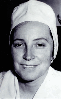
     Some time ago, a friend referred me to a Medscape list of the “Fifty Most Influential Physicians in History”. Many names on the list are familiar to physicians and medical historians, but one unfamiliar name caught my attention: Dr. Zora Janzekovic. She was noted to be a plastic surgeon from Slovenia, elevated into this group of fifty for her major contributions to the therapy of burns. Her achievement is especially remarkable considering the demanding conditions under which she worked.

     Dr. Janzekovic was born in September, 1918, in Slovenska Bistrica, Slovenia. After receiving her MD degree at the Zagreb University Medical School, she obtained specialty training in plastic surgery in Belgrade, then underwent a rapid, six-month training course in burn management in Ljubljana, Slovenia. Enough scientific exchange with the West existed, however, to ensure good training. She was assigned to run a burn unit in the city of Maribor, the second-largest city in Slovenia, situated in the northeast, where burns, especially in children, were frequent.
     On arrival, Janzekovic was greeted with almost impossible conditions. Yugoslavia was still behind the Iron Curtain and the Cold War was on. In the Maribor hospital she found that no burn unit existed. Surgical equipment, food, dressings, and medications were all scarce or absent and there was limited access to pertinent literature. Nurses and physicians had not been trained to care for burn victims, and funds to remedy conditions were meager. Children with burns came frequently, and Dr. Janzekovic was pained to witness their emaciation and suffering.
Dr. Zora Janzekovic (Wikipedia)
    But she forged ahead. She managed to acquire three rooms in the dermatology department, gradually expanding to accommodate the approximately 350 burn patients seen per year, many of them serious, and most needing painstaking care. Dressings full of pus piled up after the daily changes, creating a repugnant stench, and medications were in short supply. The staff was buckling from overwork.

     Janzekovic commandeered yet more hospital space. Equally important, she realized that the infections under the dressings were coming from the patients’ own tissues, allowing her to cut down on isolation procedures, saving time and space. She trained the nurses and acquired another physician to help.

     The major breakthrough came next. At the time, the usual practice with deep burns was to protect the burn with dressings until the superficial dead tissue demarcated from the healthier tissues beneath, then apply skin grafts. This took time, and Janzekovic wondered if one could shorten the process by simply excising the upper, apparently dead, layers of tissue only a few days after the burn and then apply grafts immediately to the denuded area. Pushed by the sheer number of patients, she tried the new approach on a few smaller burns, with success. With more experience, she honed the technique and calibrated the best timing for excisions and dressing changes. Happily, the early grafts healed more rapidly and with a minimum of scarring. Gradually she tackled larger and larger burns, many large enough to require skin grafts from other donors. Overall the new procedure saved huge amounts of time, freed up needed beds, and reduced the infection rate dramatically, saving the use of scarce antibiotics.

     Soon her colleagues from the capital, Ljubljana, came to Maribor to see for themselves, and were impressed enough to invite others from abroad. In 1968 the Burns Society of Slovenia held the Third Congress of the Yugoslav Association for Plastic and Maxillofacial Surgery in Maribor. A number of burn specialists attended, among them Douglas Jackson, from England, who tried out the method in his home city of Birmingham. Dr. Jackson, named, in 1969, to give the first Everett Idris Evans Memorial Lecture (named after the surgeon who pioneered research on fluid dynamics in burns and on radiation burns), pronounced the method successful. His opinion brushed away a good deal of skepticism and the Janzekovic method spread.

     In 1975 Janzekovic published her experience with an astounding 2,615 patients who had undergone the excision and grafting procedure. Pain was reduced, patients discharged more promptly (average stay was 14 days), aesthetic appearance improved, and contractures from scarring were relatively infrequent.

     By this time Dr. Janzekovic was well known. Between 1968 and 1984 a total of 237 burn surgeons made a pilgrimage to her clinic, and she was invited to lecture at meetings “from Los Angeles to Shanghai,” as she put it. She was chosen, in 1975, to deliver the Evans Memorial Lecture, the same lecture at which Douglas Jackson had first publicized her technique. In 2007 a new award was created by the European Club for Pediatric Burns, the Zora Janzekovic Award: “The Golden Razor”. Dr. Janzekovic was, of course, the first recipient and she received many other honors.Today her technique is standard practice in burn therapy.

     In her later years Dr. Janzekovic did research on shock in burns, though, as she said, “it was far too great a challenge for our circumstances.” She did, however, think that overheated blood might produce toxins and speculated on the use of exchange transfusions to eliminate them.

     Zora Janzekovic, after many years of tireless work and healing thousands of children, retired in her native country. She had lived through WWII, worked throughout the Cold War in communist Yugoslavia, and, finally, made the transition to the European Union. At her 90th birthday she was quoted as saying, “My life was worth having been lived.” She died in 2015 at age 96. It is fitting that she is ranked in Medscape’s 50 most influential physicians.


SOURCES:

Dr. Igor M Ravnik, in Ljubljana, kindly reviewed and helped with this essay.


Janzekovic, Z. “Once upon a time: How west discovered east”. 2008; J Plast Reconstr, Aesthet Surg 61: 240-44.


Burd, A. “Once upon a time and the timing of surgery in burns”. 2008; J Plast Reconstr Aesthet Surg 61: 237-9.


Janzekovic, Z. “A new concept in the early excision and immediate grafting of burns”. 1970; J Trauma 10: 1103-8.


Janzekovic, Z. “The burn wound from the surgical point of view”. 1975; J Trauma 15: 42-62.


Obituary. 2015; Burns 41:1374.


Barrow, R E and Herndon, D N. “History of treatments of burns”. Chapter in Herndon, D N, ed. Total Burn Care, 3rd edit. 2007; pp 1-8.


Powers, J M and Feldman, M J. “Everett Evans, nuclear war, and the birth of the civilian burn center”. 2017; Amer Coll Surg Poster Competition.