Monday, March 16, 2020


A HERO IN BURN THERAPY


     Some time ago, a friend referred me to a Medscape list of the “Fifty Most Influential Physicians in History”. Many names on the list are familiar to physicians and medical historians, but one unfamiliar name caught my attention: Dr. Zora Janzekovic. She was noted to be a plastic surgeon from Slovenia, elevated into this group of fifty for her major contributions to the therapy of burns. Her achievement is especially remarkable considering the demanding conditions under which she worked.

     Dr. Janzekovic was born in September, 1918, in Slovenska Bistrica, Slovenia. After receiving her MD degree at the Zagreb University Medical School, she obtained specialty training in plastic surgery in Belgrade, then underwent a rapid, six-month training course in burn management in Ljubljana, Slovenia. Enough scientific exchange with the West existed, however, to ensure good training. She was assigned to run a burn unit in the city of Maribor, the second-largest city in Slovenia, situated in the northeast, where burns, especially in children, were frequent.
     On arrival, Janzekovic was greeted with almost impossible conditions. Yugoslavia was still behind the Iron Curtain and the Cold War was on. In the Maribor hospital she found that no burn unit existed. Surgical equipment, food, dressings, and medications were all scarce or absent and there was limited access to pertinent literature. Nurses and physicians had not been trained to care for burn victims, and funds to remedy conditions were meager. Children with burns came frequently, and Dr. Janzekovic was pained to witness their emaciation and suffering.
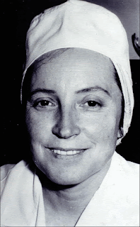
Dr. Zora Janzekovic (Wikipedia)
    But she forged ahead. She managed to acquire three rooms in the dermatology department, gradually expanding to accommodate the approximately 350 burn patients seen per year, many of them serious, and most needing painstaking care. Dressings full of pus piled up after the daily changes, creating a repugnant stench, and medications were in short supply. The staff was buckling from overwork.

     Janzekovic commandeered yet more hospital space. Equally important, she realized that the infections under the dressings were coming from the patients’ own tissues, allowing her to cut down on isolation procedures, saving time and space. She trained the nurses and acquired another physician to help.

     The major breakthrough came next. At the time, the usual practice with deep burns was to protect the burn with dressings until the superficial dead tissue demarcated from the healthier tissues beneath, then apply skin grafts. This took time, and Janzekovic wondered if one could shorten the process by simply excising the upper, apparently dead, layers of tissue only a few days after the burn and then apply grafts immediately to the denuded area. Pushed by the sheer number of patients, she tried the new approach on a few smaller burns, with success. With more experience, she honed the technique and calibrated the best timing for excisions and dressing changes. Happily, the early grafts healed more rapidly and with a minimum of scarring. Gradually she tackled larger and larger burns, many large enough to require skin grafts from other donors. Overall the new procedure saved huge amounts of time, freed up needed beds, and reduced the infection rate dramatically, saving the use of scarce antibiotics.

     Soon her colleagues from the capital, Ljubljana, came to Maribor to see for themselves, and were impressed enough to invite others from abroad. In 1968 the Burns Society of Slovenia held the Third Congress of the Yugoslav Association for Plastic and Maxillofacial Surgery in Maribor. A number of burn specialists attended, among them Douglas Jackson, from England, who tried out the method in his home city of Birmingham. Dr. Jackson, named, in 1969, to give the first Everett Idris Evans Memorial Lecture (named after the surgeon who pioneered research on fluid dynamics in burns and on radiation burns), pronounced the method successful. His opinion brushed away a good deal of skepticism and the Janzekovic method spread.

     In 1975 Janzekovic published her experience with an astounding 2,615 patients who had undergone the excision and grafting procedure. Pain was reduced, patients discharged more promptly (average stay was 14 days), aesthetic appearance improved, and contractures from scarring were relatively infrequent.

     By this time Dr. Janzekovic was well known. Between 1968 and 1984 a total of 237 burn surgeons made a pilgrimage to her clinic, and she was invited to lecture at meetings “from Los Angeles to Shanghai,” as she put it. She was chosen, in 1975, to deliver the Evans Memorial Lecture, the same lecture at which Douglas Jackson had first publicized her technique. In 2007 a new award was created by the European Club for Pediatric Burns, the Zora Janzekovic Award: “The Golden Razor”. Dr. Janzekovic was, of course, the first recipient and she received many other honors.Today her technique is standard practice in burn therapy.

     In her later years Dr. Janzekovic did research on shock in burns, though, as she said, “it was far too great a challenge for our circumstances.” She did, however, think that overheated blood might produce toxins and speculated on the use of exchange transfusions to eliminate them.

     Zora Janzekovic, after many years of tireless work and healing thousands of children, retired in her native country. She had lived through WWII, worked throughout the Cold War in communist Yugoslavia, and, finally, made the transition to the European Union. At her 90th birthday she was quoted as saying, “My life was worth having been lived.” She died in 2015 at age 96. It is fitting that she is ranked in Medscape’s 50 most influential physicians.


SOURCES:

Dr. Igor M Ravnik, in Ljubljana, kindly reviewed and helped with this essay.


Janzekovic, Z. “Once upon a time: How west discovered east”. 2008; J Plast Reconstr, Aesthet Surg 61: 240-44.


Burd, A. “Once upon a time and the timing of surgery in burns”. 2008; J Plast Reconstr Aesthet Surg 61: 237-9.


Janzekovic, Z. “A new concept in the early excision and immediate grafting of burns”. 1970; J Trauma 10: 1103-8.


Janzekovic, Z. “The burn wound from the surgical point of view”. 1975; J Trauma 15: 42-62.


Obituary. 2015; Burns 41:1374.


Barrow, R E and Herndon, D N. “History of treatments of burns”. Chapter in Herndon, D N, ed. Total Burn Care, 3rd edit. 2007; pp 1-8.


Powers, J M and Feldman, M J. “Everett Evans, nuclear war, and the birth of the civilian burn center”. 2017; Amer Coll Surg Poster Competition.

Thursday, February 13, 2020


ADOLF KUSSMAUL

          Only a rare medical student is unfamiliar with the name Kussmaul. The Kussmaul breathing of the diabetic in severe acidosis and the Kussmaul, or paradoxical, pulse of the patient with constrictive pericarditis are part of medical jargon. Adolf Kussmaul’s life and other accomplishments, though, are less well known. His career is too full to cover here but his contributions to vasculitis and gastroenterology deserve mention.
Adolf Kussmaul (Wikipedia)
     Kussmaul, the son and grandson of doctors, was born in 1822 in Baden. He received his medical education at Heidelberg, following which he studied in Vienna under illustrious pioneers such as Hebra (dermatology), Rokitansky (pathology), Semmelweiss (obstetrics), and Skoda (internal medicine and chest disease), and in Prague under Johann von Oppolzer (internal medicine). After a stint in the Army, he settled into a country practice in Kandern, southwest Germany, the birthplace of John Sutter of California Gold Rush fame. 
     In the third year of a grueling practice that included house calls at night on horseback, Kussmaul suffered a frightening, though temporary, paralysis of his legs and bladder, possibly a case of polio. After months of recovery, he decided on an academic career. At the University of Würzburg, studying under Virchow, he received his MD degree, necessary for an academic career. He started at the University at Heidelberg, teaching materia medica and forensic medicine. There he showed that rigor mortis was due to chemical changes in dying muscle and not to nerve death, as was thought. Subsequent academic moves included professorships of medicine at the University of Erlangen (1859-63), the University of Freiburg (1863-77), the University of Strassburg (1878-88). Finally, as professor emeritus of medicine, he returned to the University of Heidelberg. 
     While at Freiburg, in May of 1865, he saw a 27-year-old man admitted with pallor, severe muscular weakness, rapid pulse, and abnormal urine. Peculiar small nodules appeared on the trunk as the patient became bedridden, demented, and finally expired. At autopsy, the nodules proved to be arteries showing,
microscopically, marked nodular inflammatory changes. A second case, less severe, was diagnosed through a muscle biopsy. Kussmaul and Rudolf Maier, professor of pathology, reported the cases as a new disease, periarteritis nodosa, a malady now widely recognized. Carl von Rokitansky had previously reported the disease but, not using a microscope, failed to recognize its nature.
     Kussmaul was also a pioneer in gastroenterology. He reported, in 1869, on patients with obstruction of the stomach outlet (pyloric obstruction), presenting with upper abdominal pain, persistent vomiting, and dilated stomachs. After passing a tube into the stomach and aspirating the contents he flushed frequently with Vichywater and bicarbonate solutions. If the obstruction was due to ulcers, a few treatments, followed by a soft diet with frequent milk feedings, led to good recovery, though with occasional relapses. Those with cancer had little relief. Nasogastric tubes had been used before, especially to treat poisonings, but Kussmaul recognized their value for pyloric obstruction and for the study of stomach physiology. He also suggested the possibility of surgery as a future therapy for obstruction. Twelve years later Theodore Billroth realized the suggestion by performing the first pyloric resection. 
     Kussmaul also mentioned that he had passed a scope to see the interior of the esophagus and stomach, about which he promised to report later. The report never came, but letters have come to light shedding light on Kussmaul’s contribution.
     The idea of peering into body cavities goes back to Philipp Bozzini, a German of Italian extraction. In 1806, using a candle for a light source and employing mirrors, he looked down a short tube to see into the urethra, bladder, and rectum. The apparatus did not catch on, however. In 1853 a French surgeon, Antoine J
Desormeaux's endoscope (from his book)


Desormeaux, developed an improved instrument, which he called an “endoscope”, the first use of the word. An alcohol lamp provided light that was reflected down the scope through a mirror with a central hole, again to visualize the urethra and bladder. 
     Kussmaul was familiar with Desormeaux’s work and was inspired to design a short, functional esophagoscope. In 1867 or 8, one of Kussmaul’s assistants, curious about the anatomy of a local sword swallower, brought him to the hospital to examine his larynx. While there, Kussmaul tried out his short scope but it failed to reach the stomach. A longer rigid tube was made that the sword swallower inserted, successfully. In Kussmaul’s own words, the sword swallower showed that “…a difficulty for gastroscopy, the bending of the
Sword swallowing (photo by Bill Golladay, Wikipedia)
esophagus at the cervical level, could be overcome with the proper positioning of the head and neck” (Neumann, Hellwig). Kussmaul could see down to the junction with the stomach, beyond which he saw only foam and darkness. The sword swallower agreed to visit clinics in Basel and Zurich, teaching doctors the technique. 
     Kussmaul’s endoscope helped diagnose cancers of the esophagus and cardia and proved useful in removing foreign bodies. But the interior of the stomach remained in darkness. Rigid gastroscopy proved tricky, as well, until supplanted by the semi-flexible scope of Rudolf Schindler, and later by the far superior flexible fiberoptic instruments. 
Crown Prince Friedrich W. (Wikipedia)
     Kussmaul’s reputation as a teacher and for his skill in combining clinical investigation with bedside medicine was widespread. His prestige was such that he was called to examine the lungs of the Crown Prince Friedrich Wilhelm, who suffered from laryngeal cancer. And Kussmaul’s use of the nasogastric tube for treatment of obstructing ulcers and his introduction of rigid gastroscopy paved the way for the specialty of gastroenterology. We owe a debt to that anonymous sword swallower.

SOURCES:
Kluge, F. Adolf Kussmaul 1822-1902. 2002; Rombach Verlag.
Matteson, E L and H R. polyarteritis Nodosa and Microscopic Polyangiitis. 1998; Mayo Foundation.(A translation of the original articles)
Neumann, H A and Hellwig. Vom Schwertschlucker zur Glasfiberoptik: Die Geschichte der Gastroskopie. 2001; Urban & Vogel.
Bast, T H and Miller, W S. The Life and Time of Adolf Kussmaul. 1926, Paul B Hoeber (Reprint)
Kluge, F and Seidler, E. “Zur Erstanwendung der Ösophago-und Gastroskopie: Briefe von Adolf Kussmaul und seinern Mitarbeitern.”. Medizinhistorisches Journal  1986; 21(3): 288-307.(contains excerpts of letters by Kussmaul and colleagues)
Matteson, E and Kluge, F. “Think Clearly, Be Sincere, Act Calmly: Adolf Kussmaul (February 22, 1822 – May 28, 1902) and his Relevance to Medicine in the 21st Century.” Curr Opin Rheumatol 2003; 15: 29-34.
Desormeaux, A. De l”Endoscope et de ses Applications au Diagnostic et au Traitement des Affections de l’Urethre et de la Vessie: Lecons faites a l’Hopital Necker. 1894; J B Ballière.    

Monday, January 13, 2020


A HISTORY OF CESAREAN SECTION


       The origin of the name "cesarean section" is unknown. Three principal explanations have been suggested.                                                                                            1)   Legend says that Julius Caesar was born in this manner, hence the name "Caesarean".  There are several reasons to doubt this.  First, the mother of Julius Caesar lived for many years after his birth and in 100 B.C. the survival rate for the procedure was
Birth of Julius Caesar, 1506 French woodblock (Wellcome Library)
essentially zero.  Second, the operation, whether performed on the living or the dead, is not mentioned by any medical writer before the Middle Ages.                                                 
 2)   It may have been derived from a Roman law, supposedly created by Numa Pompilius (eighth century B.C.), ordering that the procedure be performed upon women dying in the last few weeks of pregnancy in the hope of saving the child.  This explanation then holds that the lex regia, as it was called at first, became the lex caesarea under the emperors, and the operation itself became known as the caesarean operation. The German term Kaiserschnitt reflects this derivation.                                                                                       3)   The word caesarean was possibly derived sometime in the Middle Ages from the Latin verb caedere, "to cut."  This explanation of the term caesarean seems most logical, but exactly when it was applied to the operation is uncertain. Since "section" is derived from the Latin word seco, which means "cut", the term Cesarean section seems redundant

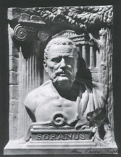
     In reference to abdominal delivery in antiquity, it is pertinent that no such operation is mentioned by Hippocrates, Galen, Celsus, Paulus, Soranus, or any other classical medical writer. If cesarean
Soranus of Ephesus (Nat Library of Medicine)
section were employed at that time, it is surprising that Soranus, whose extensive work of the second century A.D. covers all aspects of obstetrics, does not mention cesarean
section.                                          

     Several references to abdominal delivery appear in the Talmud (Jewish civil and religious law writings) between the second and sixth centuries A.D., but whether they were used in a clinical setting is doubtful. Cesarean section on the dead appears to have been practiced, however, soon after the Catholic Church gained dominance, to enable baptism of the child. Locally, two records of postmortem sections are known, one at Mission Dolores in San
C Section on expired woman (Wikipedia)
Francisco in, 1805, and another at the Santa Clara Mission, in 1825. Neither infant survived.

     The earliest cesarean section done on a living woman was reported in 1500, performed by Jacob Nufer, a castrator of pigs at Sigerehausen, Switzerland. The patient (his wife) and the baby both survived, but since the mother subsequently delivered five more children vaginally there is doubt about the report's validity. Dr. Robert Harris reported on the first known cesarean operation in the U.S. A fourteen-year-old quadroon performed it on herself in a snowbank in Nassau, New York, using an L-shaped incision, dressed by her employer. Mother and baby survived.                                                                                                                        

         Credit for the first Cesarean section performed by an American physician goes to Dr. John Lambert Richmond. Growing up impoverished, he educated himself during childhood, eventually gaining entrance to the newly organized Medical College of Ohio. Under its founder, the formidable Daniel Drake (see blog of 9/16/2019), he received his M.D. degree in 1822. He also studied to become an ordained Baptist minister. Called to a house on the
Article by Richmond (Hathi Trust)
evening of April 22, 1827, Dr. Richmond found a primigravida exhausted from a thirty-hour labor, with no cervical dilation, and with seizures (probable eclampsia). He was seven miles from home, in a storm. In Richmond's own words, "…with only a case of common pocket instruments, about one o'clock at night I commenced the cesarean section. Here I must….relate the condition of the house, which was made of logs that were green and put together not more than a week before. The crevices were not chinked, there was no chimney, nor chamber floor. The night was stormy and windy, insomuch that the assistants had to hold blankets to keep the candles from being blown out." Under the precarious candlelight, Richmond made a vertical incision, removed the placenta, and delivered a large infant that did not survive. He found no opening between uterus and vagina. The uterus was not sutured closed (custom at the time), and the abdominal wall closed in two stages. The only complication to the mother was an infected hematoma, drained. She returned to work 24 days after the operation.

     Maternal mortality rates from cesarean section in the 19th century were 85 percent or higher, with the operation done as a last resort to save the life of the mother. Dr. Harris noted that as late as 1879, cesarean section was more successful when performed by the patient herself (or ripped open by a bull's horn). He compared nine such cases from the literature, with five recoveries, to twelve cesarean sections done in New York City during the same period with only one recovery.

     The turning point came in 1882 when Max Saenger, a 28-year-old assistant to Dr. Credé at the University Clinic in Leipzig,
Max Saenger (Wikipedia)
introduced suturing of the uterine wall, using silver wires. Eight out of seventeen mothers survived their sections, remarkable for the time. New surgical techniques, antibiotics, and blood transfusions have lowered the risk dramatically since then. In 1950, D'Espo reported 1000 consecutive cesarean deliveries without a single maternal death. The frequency of the operation rose, from 5 percent of births in 1970 to 23 percent in 1985. The CDC reports a rate of 31.9 percent of births for 2018.

     The present state of cesarean section surgery enables millions of women to avoid otherwise difficult childbirths with a safe and healthy outcome.


                                                                         Michael Shea MD


SOURCES:

Creasy, R K. and Resnik, R. Maternal Fetal Medicine Principles and Practice. 1984; W.B. Saunders Company.

Cunningham, F. G, MacDonald, Paul C. and Gant, Williams, J W. Williams Obstetrics. 1989; Appleton and Lange.

Eastman, N J and Helman, L M.  Obstetrics. 1961; Appleton-Century-Crofts Inc.

Harris, Henry. California's Medical Story. 1932; Carles C. Thomas.

Speert, Harold. Obstetrics and Gynecology in America: A History. 1980; Waverly Press.

King, A G. "America's First Cesarean Section". Obstetrics and Gynecology 1971; 37(5): 797-802.

Harris, R P. "A Study and Analysis of One Hundred Cesarean Operations Performed in the United States During the Present Century and Prior to the Year 1878". Am J Med Sci 1879; 77: 43-65.


Monday, December 16, 2019


A VANISHING DISEASE:
GENERAL PARESIS


Guy de Maupassant (Wikipedia)
    On January 1, 1892, the famous French writer Guy de Maupassant, in a delirious state, pointed a revolver at himself and pulled the trigger. He heard only a click, however, as his valet, worried about his mental condition, had earlier removed the bullets. Maupassant then stabbed himself superficially and attempted to jump out a window before his valet rescued him. Committed to an asylum, Maupassant’s delusions and irascible behavior intensified and, in July of 1893, after a series of convulsions, he finally met his end. He was 43 years old. His brother had met a similar fate not long before.
    Years earlier, Maupassant proudly announced that he had syphilis, undoubtedly related to his frequent brothel visits. By the late 1880s, he was becoming irritable and complaining of pains in his stomach, head, and elsewhere, preludes to his later sufferings. His final years were almost certainly due to a late stage of syphilis, known as general paresis, characterized by delusions, dementia, weakening or paralysis of various motor functions, and eventually death.
   At the onset of the nineteenth century even the notion that brain disease could cause mental illness, rather than the other way around, was unclear. A Parisian medical student, Antoine Bayle, in his doctoral thesis of 1822, explored the issue with a path-breaking doctoral thesis. He noted that since so many autopsies in mentally ill patients had shown no brain pathology, theories had arisen placing the seat of “madness” in other organs. Bayle then described six cases from the Charenton Insane Asylum, near Paris (where the Marquis de Sade had been confined), who had progressed from delusions of grandeur to weakness, dementia, and death. In all cases, he found inflammation of the tissues covering the brain and attributed the mental disturbances to the inflammatory changes. Not long after, Bayle published on over 200 similar cases and other authors made similar reports, all boosting the theory of organic/structural causes for mental illness.
Alzheimer's drawing of involved areas in general paresis (Hist u Histopath Arbeiten v1,1904, Hathi Trust)
     Alois Alzheimer, the pathologist who described the brain changes seen in dementia, published, in 1904, detailed findings in the brains of paretics, locating the primary damage in the frontal lobes and meningeal coverings. Franz Nissl, another noted neuropathologist and a friend, published similar findings in the same journal issue. By this time general paresis accounted for about 10 to 30 percent of psychiatric admissions. A relation to syphilis was considered probable, though with no organism to point to debate was still vigorous. William Osler, in his 1892 text, acknowledged a relationship to syphilis but felt that in the U.S. the hectic pace of modern life was a major factor causing general paresis, along with alcoholism and lead poisoning. 
   Events accelerated the following year when Fritz Shaudinn finally sighted the tiny spiral-shaped organism, now called treponema pallidum, in a secondary syphilis papule. The next year the Wassermann reaction for syphilis was published, a valuable diagnostic tool. Yet another year later, Paul Ehrlich announced the first “magic bullet”, an arsenical, to treat syphilis. Finally, in 1912 Hideyo Noguchi at the Rockefeller Institute discovered the treponema in the brain and spinal cord of patients. Doubts disappeared on the cause of general paresis, though arguments on organic vs psychologic origins for other mental illnesses carried on. 
Julius Wagner-Jauregg (Wikipedia)
    Therapy remained a problem. Arsenicals were toxic and required a long, painful series of injections. Further, the drugs penetrated poorly into brain tissue. Some patients with general paresis, however, unexpectedly improved after surviving an infection, raising the question: could fever weaken the organism? An Austrian psychiatrist, Julius Wagner von Jauregg, cautiously investigated the effects of fever. Starting with tuberculin injections, he turned to malaria infection and saw improvement in a substantial number of paretics. Eventually, malaria therapy - infusing patients with the safer vivax strains - became widespread. A variety of heating devices also sprang up to duplicate the effects of malaria. The results impressed the scientific world enough to award a Nobel Pirze to Wagner-Jauregg. The treatments, however, were only a partial solution. They often produced remissions rather than cures and worked best in early cases. 
Al Capone (Wikipedia)
    A patient that suffered through the full gamut of ministrations was the notorious gangster, Al Capone. Probably contracting the disease in his teens, mental symptoms were first apparent in the Atlanta Penitentiary, where he was sent for tax evasion at age 33. Boasting extravagant exploits, he was diagnosed with megalomania due to central nervous system syphilis, confirmed with blood and spinal fluid tests. Bismuth (another heavy metal) therapy was begun. Two years later, this time in Alcatraz, his megalomania worsened and he received arsenicals and more bismuth. Near the end of his prison term he was transferred to the care of Dr. J. E. Moore, a world expert on venereal disease at Johns Hopkins Hospital. Capone was admitted under an assumed name and treated with malaria infections and possibly more heavy metal. The trustees of Johns Hopkins Hospital and a second hospital, learning the identity of their patient, both forced him to leave, so his family rented a house in Baltimore to finish treatments. To follow the results of therapy, repeated spinal taps were done under the watchful eye of Capone’s bodyguards.       
     In March 1940, Capone moved to his house on Palm Island, Florida, now a weak man, demented, a shell of his former self. He seemed to stabilize, grew fat, but did not improve mentally. In 1945 he was given penicillin, one of the first to receive it, but it did little good and in January 1947, after suffering a “stroke” he died quietly. 
    The former scourge of syphilis is today under better control and
 general paresis, the late stage of brain involvement, is rare. At an earlier time, however, general paresis was important in the evolution of psychiatric ideas and in the early development of therapy of infectious diseases before the modern “magic bullets”.

 And now to a lighter subject: HAPPY HOLIDAYS TO ALL AND A HAPPY NEW YEAR!

SOURCES:
Ropper, A H and Burrell, B D. How the Brain Lost its Mind: Sex, Hysteria, and the Riddle of Mental Illness. 2019; Avery Press.
Lerner, M G. Maupassant. 1975; George Allen & Unwin.
Hayden, D. Pox: Genius, Madness, and the Mysteries of Syphilis. 2003; Basic Books.
Solomon, H C. “General Paresis: What It Is and its Therapeutic Possibilities”. 1923; Amer J Psych 79: 623-46.
Whitrow, M. “Wagner-Jauregg and Fever Therapy” 1990; Medical History 34: 294-310.
A. J. K. “Joseph Early Moore” (obituary). 1958; Brit J Vener Dis 34: 58.
Moore, M and Solomon, H C. “Contributions of Haslam, Bayle, and Esmarch and Jessen to the History of Neurosyphilis”. 1934; Arch Neurol Psychiat. 32: 804-39.
Bergreen, L. Capone: The Man and the Era. 1994; Simon & Shuster.