Wednesday, November 13, 2019

MESMER AND ANIMAL MAGNETISM


     In 1778 a German doctor, Franz Anton Mesmer, educated at the University of Vienna Medical School, left his practice in Vienna after being shunned by a hostile medical faculty. His work as a healer using a new therapy, animal magnetism, based on an invisible fluid that permeated the universe and whose passage through the body was 
F. Anton Mesmer (Wikipedia)
essential for good health, was considered chicanery by the academic community. Chased also by rumors of inappropriate sexual behavior, he moved to Paris and opened a new practice in a fashionable area. 
   Animal magnetism did not sound outlandish to many French Enlightenment Parisians. Writings by Voltaire on Isaac Newton’s gravity, describing it as the effects of an invisible “fluid”, pervading the universe, were popular. Similar was electricity, another 
Voltaire at work (Wellcome Library)
mysterious fluid with a variety of uses, including treatment of illness. Magnetic attraction resembled gravity, and phlogiston, an invisible substance responsible for heat, still permeated scientific thought. Balloons filled with “dephlogisticated air” (hydrogen) ascended to extraordinary heights. Theories of immaterial “vitalistic” forces abounded in medical writing. Mesmer’s technique of restoring patients to health by aiding the flow of magnetic fluid through the body sounded quite reasonable to many.    
     For official recognition of his method Mesmer appealed to the French Academy of Sciences, the Royal Society of Medicine, and the Paris Faculty of Medicine. All refused to endorse him. Charles Deslon, however, a Paris Faculty member and physician to the brother of Louis VI (who later ruled France as Charles X), became a believer and adopted Mesmer’s techniques. 
         The demand for treatment was high, forcing Mesmer to conduct group therapy sessions. Patients were seated around a central tub, about a foot high, partly filled with glass and iron filings and covered in wood. Ropes around the patients’ waists transmitted the fluid from one to another, as did the linking of hands. Iron bars protruded from the barrel, angled to touch parts of the body to receive the magnetic fluid.  Attendants also touched various parts needing treatment with iron rods or pressed with their hands on the upper abdomen (and sometimes the lower part) to impart more fluid.
Patients around the barrel. No ropes in this image. Attendant on left pressing on abdomen. Notice mirrors.
(Wellcome Library)
     In individual sessions the therapist sat with his knees touching the patient’s as he pressed on various parts of the abdomen. The light was dimmed and a musical instrument, usually a glass armonica, invented by Benjamin Franklin, 
Glass harmonica. Rubbing the fingers on the edges of the
rotating wet glass discs produces the notes (Wikipedia)
produced soft, ethereal sounds. Mirrors around the room reflected back any escaping magnetic fluid. Before long subjects began trembling or shouting and progressed to seizures and other movements. Those with the most violent movements were moved to a room with padded walls. Inducing such a “crisis” was thought important to a cure. 
Another Version. The woman in the back is carried into the padded room (Wellcome Library)
(Click on image for enlargement)

     Mesmer formed a secret society, the Society of Harmony, to initiate new practitioners, for which he charged a substantial admission fee. But there were many detractors, ridiculing the methods. Especially noted was that most of the subjects experiencing crises were women.
    
Single patient (Wellcome Library)
 The controversy for and against mesmerism was so intense that King Louis XVI established a royal commission to investigate. It consisted of five members of the Academy of Sciences and four from the Faculty of Medicine. Prominent members included the chemist Lavoisier, Bailly (a famous astronomer), Dr. Guillotin (for whom the beheading instrument was named), and the famous botanist Laurent de Jussieu. Benjamin Franklin, the most famous of all, headed the commission. A second commission filing a secret report included five from the Royal Society of Medicine. 
Benjamin Franklin, by Joseph Duplessis
(Wikipedia)
     Mesmer refused to cooperate with the commission unless they considered long-term results, which they declined to do. Mesmer and his chief disciple, Deslon, however, had separated. Deslon, already magnetized, knew the technique and had established his own practice, so the commission turned to him to magnetize subjects. 
     The commissioners understood that “cures” could be spontaneous, unrelated to treatment, and focused their attention on establishing whether the “fluid” existed or could be detected by “sensitive” patients. Both commission members and patients were tested. The commissioners, when magnetized, felt nothing. Subjects, when blindfolded and asked to touch objects that might or might not be magnetized, failed to detect anything accurately. 
    After many trials, often at Benjamin Franklin’s residence in Passy, a Paris suburb, the commission concluded that the “cures” could only be attributed to patient suggestion and that there was no evidence of a mysterious “fluid”. The commission wrote “…that the imagination without the magnetism produces convulsions, and that the magnetism without the imagination produces nothing; they have concluded…that the existence of the fluid is absolutely destitute of proof, that the fluid having no existence can consequently have no use…” The second, “secret”, report for the King’s eyes, by the Royal Academy of Medicine, suggested that the various touchings during the process may have awakened sexual desires in the mostly female subjects, and indicated that women were more susceptible to suggestion. Definitely a male point of view. 
   After the report Mesmer’s influence waned. As the revolution approached, he traveled to Switzerland and Germany, partly to visit family, and ended his days in obscurity.
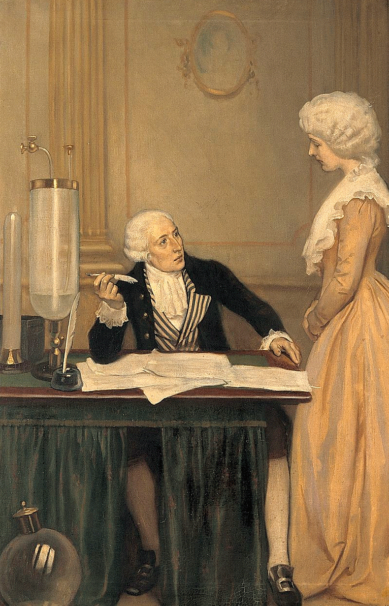
Antoine Lavoisier and his wife
(Wikipedia)
  The ensuing revolution in 1789 changed France and French medicine forever. Both Lavoisier and Bailly  perished under the guillotine. Dr. Guillotin was imprisoned but eventually released. The Marquis de Lafayette, another devotee, tried to introduce mesmerism to the U.S. but Thomas Jefferson opposed it and it never grew deep roots. The term “mesmerized” took on a milder connotation. Mesmer’s work, however, was a step toward the eventual recognition of the subconscious.

  
  SOURCES:
Darnton, Robert. Mesmerism and the End of the Enlightenment in France. 1968; Harvard Univ Press. 
Buranelli, Vincent. The Wizard from Vienna. 1975; Coward, McCann & Geoghegan.
Animal Magnetism: Report of Dr. Franklin and Other Commissioners (English Translation) 1837; H. Perkins, Philadelphia.
Huth, E J. “Benjamin Franklin’s Place in the History of Medicine”. 2006; James Lind Library Bulletin, available at: https://www.jameslindlibrary.org/articles/benjamin-franklins-1706-1790-place-in-the-history-of-medicine/
The International Journal of Clinical and Experimental Hypnosis, v 50 (4), 2002, devoted the entire issue to Franklin and Mesmer.
Lopez, C. “Franklin and Mesmer: An Encounter”. 1993; Yale J Biol Med 66: 325-31.


Tuesday, October 15, 2019


FROM GOLD MINER TO SURGERY PROFESSOR:

THE LIFE OF HUGH TOLAND


          Last month’s blog featured Transylvania University as the first medical school in the west. One of their graduates, it happens, went on to be the founder of the University of California San Francisco Medical School. His name was Hugh Huger Toland.

     Hugh Toland entered the world in Guilder’s Creek, South Carolina, in 1806. His father, a prosperous planter, noting Hugh’s early interest in natural history, arranged an apprenticeship with a local practitioner, after which Hugh enrolled in the Transylvania U. Medical School. After receiving his MD degree in 1828, graduating at the top of his class, Toland practiced in a small town in South Carolina. Bills were seldom paid, and to supplement his income he served as an itinerant healer, traveling to various rural areas. On these travels he taught himself French, meanwhile earning sufficient money to travel to Paris. There he studied
Hugh Huger Toland (Wikipedia)
medicine, and especially surgery, under luminaries such as Guillaume Dupuytren, Jacques Lisfranc, Velpeau, and others.

     Fortified with invaluable experience he returned after 2½ years, opened a practice in Columbia, S.C., and married. By 1852, with civil war brewing and attracted by California gold fever, he set out for California in a Conestoga wagon, accompanied by a second wife (his first wife died). He made it overland from Independence, MO in 76 days, said to be a record. Sadly, his second wife died of dysentery or cholera three days after arrival. He bought a mining claim at Mokelumne Hill, Calaveras County, and mined gold for a few months but decided that mining was not for him. Returning to San Francisco, he opened a practice, at first in a partnership, then solo. His practice prospered as he saw up to 100 patients a day, patients who took their prescriptions to the adjoining drugstore that he owned. He also carried on a mail order drug business with numerous miners who mailed in their symptoms. Wells Fargo agents collected the fees. His surgical training helped him to achieve a reputation as an excellent surgeon, aided no doubt by the recent introduction of anesthesia. He married a third time, saved money (he was generally frugal), bought considerable land, and grew wealthy.

     San Francisco in the 1850s was corrupt and violent. In 1856 a former banker turned newspaper editor, James King of William (there were three James Kings in town and this one was the son of William King) used his paper to expose James Casey, a former inmate of Sing Sing Prison who had been elected to the Board of Supervisors through a stuffed ballot box. Casey responded to the exposé by shooting King in the upper chest. Two young surgeons, one named R. Beverly Cole (mainly practicing OB-GYN), rushed to help. Thinking the subclavian artery had been severed they
R. Beverly Cole (from Physicians and
Surgeons of America, 1896)
inserted a sponge to stop the bleeding. Later other physicians,
including Toland, appeared. Cole soon felt that the sponge could be safely removed, to avoid suppuration. But Toland, 23 years older than Cole and now a respected, experienced surgeon, felt it should remain (as did others) because of an absent pulse in the adjacent arm, and remain it did. King died of sepsis about a week later. Casey, meanwhile, had been convicted and hung by a rapidly assembled Vigilance Committee.

     At autopsy the subclavian artery was found intact. During a State Medical Society meeting Cole labeled Toland’s retention of the sponge malpractice and made similar remarks at the trial of an accomplice of Casey. Toland’s decision was considered legitimate but, needless to say, Cole and Toland were not friends.

     Three years later Dr. Elias Samuel Cooper formed the Medical Department of the University of the Pacific, the first San Francisco medical school, naming Cole as professor of obstetrics. Cooper died in a few years (1862) and his nephew, Levi Cooper Lane, could not sustain the school. Toland, in 1864, flush with ample money from his practice, opened his own medical school, inviting the U of P faculty, except Cole, to join. The school, on
Stockton St. near Chestnut St, used the nearby City and County Hospital for teaching purposes. It grew rapidly, but when a new dean arrived the old U of P faculty and most of the students left t0 form another school, one that eventually became the Stanford University School of Medicine. Toland, needing help, bit his tongue and asked Cole, now a physician influential in City affairs, to assume deanship of the Toland School.

     In 1869 the new University of California opened its doors. Toland wanted an affiliation and Cole was instrumental in
Toland Hall, University of California Medical Department
(from Annual Announcement of Lectures at Toland Hall, 1875.
Internet Archives)
negotiations with the University. Though the school was a gift from Toland, he had to drop the Toland name in return for a lecture hall in his name and a professorship of surgery.


      Students rated Toland’s lectures and bedside rounds highly, and in 1877 the lectures were published. Of interest is that they do not mention Lister’s antiseptic approach, but a former student indicates that he “insisted on the importance of absolute cleanliness”. He was
Rhinoplasty, from Toland's Textbook of Surgery, 1877,
before and after pictures. (Hathi Trust)
particularly known for bone surgery. He also excelled in vascular surgery (mainly ligations), bladder stone removals, and he was familiar with flaps for facial plastic surgery. Usually he worked seven days a week, devoting little time to social activities.

     Toland continued in active practice until February 1880, when, as he was walking downstairs to go to work, he fainted, fell, struck his head, and expired shortly thereafter. Reflecting the large number of devoted patients he served, the funeral was said to be “the largest ever held in San Francisco.”


SOURCES:

Gilcreest, EL. “Hugh Huger Toland”. 1938; Calif and West Med 48 (4); 263-6 and (5): 350-3.

Gardner, FT. “The Little Acorn: Hugh Huger Toland, 1806-1880”. 1950; Bull Hist Med 24: 61-9.

Toland, HH. Lectures on Surgery. 1877; Lindsay and Blakiston, Philadelphia.


McLean, R. “Hugh Toland, 1806-1880.” Unpublished manuscript, Archives of UCSF Library.

Toland, HH. “Report of the Committee on Surgery”. Trans Med Soc State of Calif 1874-5; 5 (ns): 45-50.

Lyman, GD. “The Sponge” 1928; AnnMed Hist 10: 460-79.

Gardner, FT. “King Cole of California”. 1940; Ann Med Hist 2 (3rd series): 245-58, 319-47, 432-42.


Monday, September 16, 2019


THE FIRST WESTERN MEDICAL SCHOOL


     In the decades after the American Revolution the “west” meant anything on the Pacific side of the Appalachian Mountains. A forested area of western Virginia was called the Transylvanian Colony, near which, in Lexington, a small Transylvania Seminary served as an educational center. In 1792 most of the area moved into the newly-formed state of Kentucky and 7 years later the Seminary merged with a rival school, the Kentucky Academy, to become Transylvania University. It included a law and medical
Transylvania Colony circa 1792 (Wikipedia)
school and earned Lexington the label “the Athens of the West”. Henry Clay, later a well-known senator, taught in the law school. The medical school had a faculty of two, Dr. Frederick Ridgely and Dr. Samuel Brown. Ridgely had been schooled in Philadelphia and served in the Revolutionary Army, while Brown had studied medicine at Edinburgh. Instruction was mainly out of their homes, with no anatomical dissections and few medical students. In 1815, however, a major overhaul began, led by the prominent and feisty surgeon, Benjamin Winslow Dudley.  
Benjamin Dudley (Wikipedia)
  

     Dudley had been apprenticed to Dr.  Ridgely. He then earned an MD degree at the University of Pennsylvania Medical School after 2 winter seasons of lectures (standard at the time). There he met 2 men important to him in later life: Daniel Drake and William Richardson. He headed for  Paris where he studied surgery with Dominique Jean Larrey, the famous surgeon who served Napoleon’s army. In London he studied with John Abernathy and Astley Cooper, both excellent surgeons, and passed the exams for admission to the Royal College of Surgeons. He was the most educated surgeon in the “west” and was appointed professor of anatomy and surgery at the Transylvania Medical School. He became famous for doing over 200 lithotomies with about a 2% mortality, unheard of at the time and possibly related to his habit of washing all his instruments with boiled water. He also published on numerous other conditions.

     Dudley recruited other talented faculty. The most important catch was Daniel Drake, appointed professor of materia medica. Drake, born in New Jersey, grew up in a log cabin in the small
Daniel drake (Wikipedia)
settlement of Mays Lick, Kentucky, where danger of Indian attacks still lurked. At age 15, barely literate, the boy was apprenticed to a Cincinnati practitioner, Dr. Goforth, who granted him a “diploma” after 4 years. Goforth was an enlightened physician for his time and introduced vaccination to Cincinnati. Drake then studied one year (1805-6) at the U of Penn under Benjamin Rush, completing his second year in 1816, gaining him an MD degree. He was bookish, read widely, and built a successful practice in Cincinnati. When Dudley offered him a professorship in materia medica and medical botany in Lexington he took it. He enjoyed teaching and the students consistently rated his lectures as the best.

     A third faculty member appointed was William Richardson, an acquaintance from U of Penn days. He had only completed one
William Richardson (from Hathi Trust)


semester at Penn and practiced obstetrics. He became professor of midwifery at Transylvania U. Two other new appointments, James Overton as Professor of “Theory and Practice” and James Blythe as Professor of Chemistry, filled out the new faculty by 1817.

     There was strife from the start. In 1818 Dudley and Richardson fought a duel. Dudley shot Richardson in the groin, severing an artery. Dudley then saved him by pressing on the artery until it could be tied. They continued to work together but it is unclear how harmoniously.

     Drake, to escape tensions, moved back to Cincinnati after one year, resumed his practice, and founded his own medical school, the Medical College of Ohio. He studied and wrote on diseases of the Ohio Valley and later taught once more at Transylvania U. He is regarded as a founder of western medicine.

     In the same year as the duel, the Kentucky legislature appropriated money for an upgrade of Transylvania U. The trustees hired Horace Holley, a Unitarian minister from Boston, as president. Holley quickly transformed the University. He enlisted Charles Caldwell from the University of Pennsylvania as dean of the medical school, who traveled to Europe to acquire scientific apparatus and especially books, the start of a fine library. There being no separate medical building, Dudley built an amphitheater onto his own house for dissections and surgical instruction. Cadavers were procured by graverobbing or sometimes by the use of deceased slaves. Other classes were held in the main building until a separate building was erected in 1839. In 1828 the respected quarterly, The Transylvania Journal of Medicine, was launched.

     The school soon began to be affected by the steamboat age. Louisville and Cincinnati, both on the Ohio River, grew rapidly as Lexington, farther from navigable rivers, remained more static. A medical school opened in Louisville, galvanizing the Lexington legislature to appropriate more money. A new medical building went up and
(from The History of the Medical Department of Transylvania University)
another team went to Europe for more books and equipment. The new library was praised as the finest in the west and among the finest anywhere. Nathan Smith (see blog of 8/12/16) and Elisha Bartlett, famous names at the time, taught there and the school flourished through the 1840s. But the schools in Louisville and Cincinnati gained in size and in 1850 several of the Transylvania faculty opened a new medical school, the Kentucky Medical School, in Louisville, to compete. Eventually, in 1860, the Transylvania medical school closed, and the new one in Louisville merged with the University of Louisville in 1908. Transylvania U eventually became the University of Kentucky.

     The Transylvania U medical school had a short but illustrious life, easily comparable to eastern schools and responsible for training most of the practitioners of the early west.


SOURCES CONSULTED:

Wright, John D. Transylvania: Tutor to the West. 1975; University Press of Kentucky.

Juettner, Otto. Daniel Drake and his Followers. 1909; Harvey Publishing Co.

Wright, James R. “Early History of Transylvania Medical College” 2019; Clin Anat 32: 489-500.

Peter, Robert. The History of the Medical Department of Transylvania University. 1905; John P Morton & Co, Louisville.

Flexner, James Thomas. “Genius on the Ohio: Daniel Drake”, chapter in Doctors on Horseback. 1937; Viking Press. pp 165-234.


Tuesday, August 13, 2019

A CONTROVERSIAL NOBEL PRIZE

     Few would dispute that Nobel Prizes in the sciences are seldom mistakenly awarded. Occasionally, but only occasionally, the prize has been viewed later as inappropriate. One such instance was the award made to Dr. Johannes A. G. Fibiger, in 1926, for his work showing that intestinal worms could cause cancer. Fibiger’s conclusions eventually proved invalid but his research and life story are worth reviewing.
     Fibiger was born in Denmark in 1867. He received his medical degree from the University of Copenhagen and later studied
Johannes Fibiger (Wikipedia and National
Library of Medicine)
bacteriology in Germany. At the age of 33 he was named director of the Institute of Pathological Anatomy at the University of Copenhagen, a post he held until his death 28 years later.
     In 1907 Fibiger noticed stomach tumors in wild rats and on sectioning found small roundworms inside the growths. He learned that the worms, which he named Spiroptera carcinoma (now called Gongylonema neoplastica), were common in cockroaches, existing as a larval form in their muscles. He fed cockroaches infected with larvae to rats and produced stomach tumors, though feeding adult worms produced none. He reported the results in 1913 and the work was considered a breakthrough. However, criticisms of the report came quickly, centered mainly around whether the histology was consistent with cancer. Fibiger reported metastases in some rats but again the histology was disputed. Other workers could not easily duplicate the findings.
     In 1926 Fibiger and Katsusaburo Yamagiwa (who had induced cancer by painting coal tar on rabbit ears) were considered jointly for the Nobel Prize in medicine. No decision could be reached, however, and the prize was postponed. It was finally given to
Nobel Prize medal (Wikipedia)
Fibiger alone in 1927. The 1927 prize was awarded at the same time to Julius Wagner-Jauregg for his discovery of the helpful effect of malaria in treating tertiary syphilis (also considered a mistake by some). The staff at the Karolinska Institute, that awards the prize, commented on Fibiger’s work that “It was thus shown authoritatively not that cancer is always caused by a worm, but that it can be provoked by an external stimulus. For this reason alone, the discovery was of incalculable importance.” In his acceptance speech Fibiger acknowledged that helminthes occupied “only a modest place among the causes of neoplasms among humans”, adding that physical and chemical influences and “endogenic and exogenic” factors all played a role.
     How did the Nobel committee make the decision to award Fibiger the prize? Several seasoned pathologists had agreed with Fibiger’s histologic diagnosis of cancer, though today it would be considered hyperplasia (non-cancerous excessive growth). Cancer was known to be more common in certain occupations, such as chimney sweepers, chemical factory workers, and after radiation exposure but laboratory attempts at cancer production had generally been unsuccessful, Katsusaburo’s work being an exception. Thus the work was considered a step forward. Doubters and critics of Fibiger’s work remained, however.
     In 1937 it was shown that vitamin A-deficient diets could lead to similar changes in stomach linings. Fibiger’s rats were probably fed a vitamin A-deficient diet of bread (made without milk or egg) and water. In 1952, Hitchcock and Bell repeated
Plate from Fibiger's study of rat tumors (Hathi
Trust.  Click image to enlarge)

Fibiger’s experiments using rats on and off a vitamin A-deficient diet and produced stomach lesions resembling those of Fibiger’s animals (the original slides were studied). They concluded that vitamin A deficiency was necessary for the hyperplasia-inducing effect of the worms.
     Fibiger made another contribution to medicine, perhaps a more important one from today’s perspective. In 1896-7, before his cancer work and while working as a junior physician in Copenhagen, he carried out a clinical trial of an antiserum against diphtheria developed by Behring and Kitasato. (Fibiger had studied earlier under Koch and Behring.) Antiserum trials were successful in animals, but in humans had yielded uncertain results. Fibiger devised a randomized trial whereby children admitted on alternate days were treated alternately with conventional means (painting the throat with silver nitrate or tar oil) or conventional means plus antiserum. The study lasted one year. Eight of 239 in the serum group and 30 out of 245 in the control group died (the epidemic had a fairly low mortality rate). 60% developed serum sickness. The X2 test, developed two years later, would have shown a p-value of 0.0003.
     This is believed to be the first controlled therapeutic trial using randomization. Fibiger consciously sought to “eliminate completely the play of chance and the influence of subjective judgment”. He realized the value of large numbers of subjects to eliminate chance variations and the value of long duration of study to eliminate seasonal variations in severity/mortality.
     The study had one immediate effect - an immediate boost in demand for serum. Surprisingly, the technique of random allocation did not catch on rapidly. Excepting a few small trials, generally with scanty details on technique, it took a major British Medical Research Council trial of streptomycin against tuberculosis in 1948 to put the technique on the map.
     Fibiger died the year after his prize was awarded. He had given up working with parasites and was working on coal tar painting to induce cancer. He was a member of numerous prestigious societies, was well-regarded by his colleagues as a careful researcher, and probably would have changed his mind about the stomach tumors as new evidence accumulated. He worked at a time when knowledge of cancer and histology was still patchy. Unfortunately he entered a blind alley, a hazard threatening many an investigator.

SOURCES
Stolley, PD and Lasky, T. “Johannes Fibiger and His Nobel Prize for the Hyposthesis that a Worm Causes Stomach Cancer”. 1992; Ann Internal Med 116: 765.

Bullock, FD and Rohdenburg, GL. “Experimental ‘Carcinomata’ of Animals and their Relation to True Malignant Tumors. 1917; J Cancer Research 3: 227.

 Fibiger, J. “On Spiroptera Carcinomata and their Relation to True Malignant Tumors; with Some Remarks on Cancer Age” 1919; J Cancer research 4: 367.

 Hitchcock, CR and Bell, ET. “Studies on the Nematode Parasite, Gongylonema neoplasticum (Spiroptera neoplasticum), and Avitaminosis A in the Forestomach of Rats: Comparison with Fibiger’s Results. 1952; J National Cancer Institute 12: 1345.

Hróbjartsson, A, et al. “The Controlled Clinical Trial Turns 100 Years: Fibiger’s Trial of Serum Treatment of Diphtheria” 1998; BMJ 317: 1243.

Nobel web site: https://www.nobelprize.org/prizes/medicine/1926/fibiger/biographical/. His biography and acceptance speech are both there.


Monday, July 15, 2019

VIEWING BONES THROUGH TELESCOPES
   by Roy Meals MD

     Probably primitive man's curiosity markedly increased soon after he stood up and started walking on just his feet. He could both peek into caves and drop back onto all fours to peer down badger holes. Looking into his family’s mouths and ears soon followed. Many generations later his progeny developed metal tubes and glimpsed human interiors through all of our natural orifices. Lighting, however, was always an issue, and the torch that satisfactorily illuminated the cave was poorly accepted by early patients in the proctology clinic. FIGURE 1


     This changed in 1879 with Edison’s invention of the incandescent light bulb. Just seven years later, two German doctors were lighting up bladders with a tiny bulb on the end of a steel tube through which they squinted. Heat from the bulb and risk of breakage, however, posed problems. Nonetheless, enterprising doctors began poking holes in the skin and exploring the bladder, abdomen, and chest with lighted tubes. In 1912, Severin Nordentoft, a Danish doctor, extended this concept to the knee and coined the word “arthroscopy” (joint-view). Multiple investigators from the world around then refined and continue to refine the technique.  
     Prior to antibiotics, tuberculosis, especially in the knee, occupied much of orthopedists’ time. This was particularly so in Japan, where squatting and kneeling have long been cultural imperatives. In 1918 Doctor Kenji Takagi began using a bladder scope to examine tuberculous knees. His idea was to develop early treatment that would preclude the awkward outcome of an entirely stiff knee. Over the next 20 years he designed and tested 12 versions of arthroscopes that were progressively smaller in diameter and that incorporated better optical systems. None of them, however, were entirely practical. 

     After World War II, Takagi’s student, Masaki Watanabe, took up the banner and continued to make design improvements. In 1957, Watanabe presented a color movie describing his work, first to an international orthopedic meeting in Spain and then to major European and North American orthopedic groups on his way home to Japan. The response was tepid at best.

     Undaunted, Watanabe pressed on. The twenty-first version finally provided an adequate view and good focus even though it necessitated grinding each lens by hand. By 1958 this version became the world’s first production arthroscope, but breakage of the incandescent bulb on the end of the tube continued to be problematic. Watanabe began to receive international visitors interested in learning his technique; but when they returned home, began using it, and reported their results, collegial criticism, even ridicule, prevailed.

     In 1967 the twenty-second version, for the first time, incorporated a novel fiber optic cable. Now the hot, fragile light bulb could be 6 - 10 feet away from the operative field and transmit “cold light” into the knee joint via thousands of bundled glass threads.

     Watanabe developed at least three more versions to further address the conflicting goals of better illumination and visualization vs. smaller diameter scopes that could probe the deepest recesses of small joints. His final version was less than 1/12th of an inch in diameter—about the diameter of a coat hanger wire.  Later came miniaturized television cameras that could be attached to the arthroscope. A video monitor in the operating room displayed the images. Now residents, nurses, and students no longer had to stare at the back of the surgeon’s head as he squinted into an eyepiece attached to a narrow tube. Patients, when awake, could watch too, and a video recording of the event later allowed their families untold hours of viewing pleasure. Well, maybe minutes.

     Along with further advances in arthroscopic instrument and in scope design, international interest began to grow. At first, every procedure was merely diagnostic and was followed immediately by a large incision and exploration of the joint under direct vision to treat whatever pathology the arthroscope had revealed.

     Tiny nippers and shavers, first manual and then also powered, began to allow for arthroscopic treatment as well as diagnosis. Current techniques and instruments even allow the surgeon to place and tie sutures inside a joint. Such minimally invasive surgery allows for faster and more complete rehabilitation. Because the knee joint is large, the innovations started there, but now orthopedists also routinely apply these techniques to the shoulder, elbow, wrist, hip, and ankle joints.      Undoubtedly our caveman ancestors, torches and clubs in hand, would be pleased to know where their curiosity for peering into holes has led. FIGURE 2


Sources:
Jackson RW: A history of arthroscopy. Arthroscopy 2010; 26 (1): 91–103
Spaner SJ, Warnock GL: A brief history of endoscopy, laparoscopy, and laparoscopic surgery. J Laparoendosc Adv Surg Tech A. 1997;7(6):369-73.
Treuting R: Minimally invasive orthopedic surgery: arthroscopy. Ochsner J 2000; 2(3): 158–163.


Thursday, June 13, 2019

ALBERT NEISSER AND HUMAN EXPERIMENTATION


     Medical experimentation on humans has a long and sometimes depressing history, brought into high relief by experiences in Germany and Japan during WWII. After the war numerous papers, declarations, and laws emerged, coalescing into a more definitive approach to human experimentation. Long before the war, though, the case of Albert Neisser aroused great interest in Germany and gave birth to what is thought to be the first governmental guidelines for human experimentation.
     Albert Neisser was born in 1855 in a small Silesian town to a physician father. He studied medicine at the pretigious Breslau University Medical School (where Robert Koch had given his demonstration of the cause of anthrax in 1876, probably while Neisser was there). After obtaining his MD degree he joined the Breslau University Dermatology Clinic. 
Albert Neisser (National Library of Medicine)
     Dermatology, in those days, dealt with venereal diseases, including gonorrhea. Bacteriology, the new, popular science, induced Neisser to seek a bacterial cause for gonorrhea. He had learned microbiology at Breslau and was familiar with Abbe’s condenser and oil immersion techniques, new in the 1870s, that improved the resolution of microscopic images. At the age of 24, using the latest microscopic equipment, he discovered, in urethral discharges, the paired cocci that bear his name (Neisseria gonorrhea), and cultured them shortly afterward – an important discovery.
     After a trip to Norway to work with Armauer Hansen on leprosy, that ended in a priority dispute, Neisser returned to Germany and rose to head the Breslau dermatology clinic after his chief died. Neisser was impressed with the recent demonstrations by Behring and Kitasato of the healing effects of antiserum in cases of diphtheria. He wondered if serum from people with syphilis, made cell-free for purity, could provide similar “passive immunization” against the disease. First he injected subcutaneously serum from a patient with early syphilis into four female patients, aged 10 to 24. None developed syphilis. Next, using cell-free serum from patients with syphilis in various stages, he injected up to 30cc intravenously into four prostitutes, aged 17 to 20. They all developed syphilis. None had given informed consent.
     News of the experiments reached the press, creating an uproar. Neisser wrote a statement defending his work, suggesting also that that the latter four women contracted syphilis because they were prostitutes and not because of his serum injections. Similar experiments were done by other physicians, and he was supported by the majority of his colleagues though one, the psychiatrist Albert Moll, who was writing a book on physicians’ ethics, spoke out against him. Neisser was fined by the Royal Disciplinary Court - because he had not obtained the patients’ consent, not because of questionable science.
     The Prussian Parliament took up the case and sought an opinion from the Scientific Medical Office of Health. Rudolf Virchow, Emil von Behring, and other prominent physicians were on the panel. Lawyers were also consulted. In 1900 the Minister for Religious, Educational, and Medical affairs issued a directive. All medical interventions other than for diagnosis or treatment were prohibited if the subject was a minor or not competent, or if consent was not obtained after proper explanation of possible negative consequences. All research interventions had to be authorized by the medical director of the institution and all details had to be documented in the medical record. The directive was not legally binding.
     Evidently similar cases subsequently came to light and in 1931, in the context of an overall reform of criminal law in Germany, the national government issued “guidelines for new therapy and human experimentation”. These guidelines distinguished between experimental new treatments and experiments intended to extend knowledge but without therapeutic benefit. In both cases minors and incompetents were excluded, and consent from a properly informed and autonomous subject was required for any experiment. The only exception was if a new treatment were desired in urgent cases and immediate consent was impossible. Experimentation on dying patients was prohibited and animal experimentation should precede that on humans. Exploitation of financial or social needs was prohibited. The physician’s special responsibilities in clinical trials should be emphasized in medical teaching. A form of institutional review board was discussed but the review and oversight function was left with the medical director. The complete guidelines (translated) are found at: file://localhost/Users/john/Documents/ history of medicine & science/ethics,GERMAN GUIDELINES ON HUMAN EXPERIMENTATION 1931.webarchive. They are believed to be the first such guidelines issued by a government. They were not annulled during the Nazi era but were certainly ignored. 
Fritz Schaudinn, co-discoverer with E. Hoffmann
of Treponema Pallidum
(National Library of Medicine)
     Neisser turned to monkeys for further syphilis research and moved to Java where monkeys were abundant. He clarified several aspects of syphilis, including showing 
Erich Hoffmann, co-discoverer of Treponema
Pallidum
 (Wellcome Library)
inability to arrest the disease by removal of the primary chancre (a common belief at the time), or to immunize monkeys. He published a book summarizing the work. While he was in Java the causative Treponema pallidum was discovered and on his return he and his assistants worked with August Wasserman to develop the Wasserman test (1906). 
August Wassermann (Wikipedia)
     Neisser took an increasing interest in public health aspects of venereal disease, advocating public health clinics and regulation of prostitution, all at a time when these subjects were seldom discussed in public. He died in 1931 from complications after surgery for bladder stone.
      Neisser’s career encompassed much of modern knowledge of syphilis. His one irresponsible experiment brought the issue of human experimentation into the German public arena, where the first governmental guidelines for human experimentation were formulated, well before similar rules existed in the United States.

 SOURCES:
Vollmann, J and Winau, R. “Informed Consent in Human Experimentation before the Nuremberg Code” 1996: BMJ 313: 1445-7

Oriel, J. “Eminent Venereologists, 1. Albert Neisser” 1989; Genitourin Med 65: 229-234.

Ligon, B L. “Albert Ludwig Sigesmund Neisser: Discoverer of the Cause
of Gonorrhea” 2005; Semin Pediatr Infect Dis 16: 336-41.

 Benedek, T G. “Case Neisser: Experimental Design, the Beginnings of Immunology, and Informed Consent” 2014; Perspect Biol Med 57(2): 249-67.