Saturday, April 9, 2016

Alexander Hamilton to Rockefeller Center:
David Hosack MD

           Tension gripped the air as two men faced each other in a tragic duel. Alexander Hamilton had just arrived at a lonely spot amongst dense cedar trees on the New Jersey shore where Aaron Burr was awaiting him, early July, 1804. At a prearranged signal Burr fired, wounding Hamilton in the right side, the bullet lodging in his lumbar spine. Hamilton sank to the ground, partly supported by his second who shouted for medical help. Doctor David Hosack, waiting at the shore by the boat that had ferried them over, scrambled up to the site. The devastating scene is best
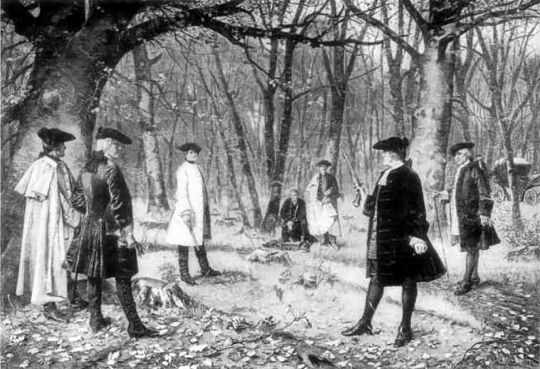
Scene of the Duel (Wikipedia)
recounted in his own words: “His countenance of death I shall never forget. He had at that instant just strength to say, ‘This is a mortal wound, Doctor', when he sunk away and became to all appearance lifeless…. His pulse was not to be felt; his respiration was entirely suspended: and upon laying my hand on his heart, and perceiving no motion there, I considered him irrecoverably gone…” (from a letter to the editor of The New York Evening Post). Hosack liberally applied spirits of hartshorn (an ammonia solution distilled from the horns and hooves of deer, later called smelling salts). On the skiff back to New York Hamilton regained consciousness, started breathing more normally and his pulse felt stronger. He complained that he had no sensation in his legs. He was placed in the house of a friend and given laudanum and other pain relievers. Hosack called in consulting surgeons, but none had more to offer and Hamilton expired in pain the following afternoon, a great loss to the young nation.

     Who was Dr. Hosack? He was, in fact, an important figure in the early history of medicine in New York. As physician to Hamilton’s family, he had watched over one son severely ill with scarlet fever, and had attended another son dying from wounds inflicted in a duel 3 years earlier. He had also been consulted by Burr in the past.
      David Hosack was born in 1769 on Manhattan, where he spent his childhood during the Revolution while British troops roamed the Island. He attended Columbia College (the name “King’s” College had been dropped) and the College of New Jersey at Princeton. He studied medicine at the Medical College of the University of Pennsylvania, where he befriended Benjamin Rush, followed by nine months at the University of Edinburgh and a year in London. In London he studied botany at the Linnaean herbarium under James Edward Smith and became a dedicated plant lover.
David Hosack by Rembrandt Peale (Wikipedia)
     Back in New York Hosack entered private practice and did well. He was on the faculty of the Columbia Medical School, and later partnered in practice with Dr. Samuel Bard, perhaps the best-known physician in New York. In 1795 and 1798 he and Bard worked through yellow fever epidemics, both contracting yellow fever in the process. As treatment he employed Glauber’s salts (sodium sulfate, a moderate laxative), bathed the patient with vinegar and cool water, and applied warm blankets while feeding liquids (called the “stove-room technique” by some). Aware of Benjamin Rush’s regimen of bleeding and violent purges he tried it in the 1798 epidemic. But after losing 40 patients he reverted to his milder method, with better results. The milder treatment also brought him many patients. He was a cofounder of the Medical and Philosophical Register, a respected medical journal, was the first in the U.S. to ligate the femoral artery for aneurysm, and innovated treatment of hydrocele by injection. He wrote many medical essays, and his practice included most of the luminaries of New York Society.
     Botany was little taught in New York and there were no large herbaria in the country. Hosack saw the need and purchased twenty acres of land between what is now 47th and 51st Streets to create the Elgin Botanic Garden (named after his father’s home town in Scotland). He poured his heart, and his money, into the garden,
Engraving, Elgin Garden. (Medical Repository 1810)
 ordering plants from around the world, quickly becoming recognized as an expert botanist. As Professor of Botany and Materia Medica at the College of Physicians and Surgeons he regularly took his medical students through the garden, teaching. 
     Hosack fostered the development of arts in New York. He was a founder of the New York Historical Society, the NY Academy of the Arts, and supported other organizations. He was an outgoing person, enjoying his students as well as social company. In 1825 he married a wealthy woman (2 other wives had died) allowing him to purchase a large estate at Hyde Park. There he built a large garden and entertained in style, hosting many notables of the day and giving up most of his practice. He died after a stroke in 1835.
     The Elgin Garden of his dreams had long before proved too expensive to maintain and he sold it to New York State in 1811. It was transferred to the College of Physicians and Surgeons, then to Columbia College. John D. Rockefeller Jr. leased the land in 1928 and built the giant Rockefeller Center on the site. Columbia sold it only in 1985. Now, strolling in Rockefeller Center on the way to the ice rink one can see a small plaque commemorating Dr. Hosack, part of which reads, “In memory of david hosack, 1769-1835, BOTANIST, PHYSICIAN, MAN OF SCIENCE AND CITIZEN OF THE WORLD”.
    
Sources:
   Robbins, C.C. David Hosack: Citizen of New York. 1964
   Hamilton, A.M.: The Intimate Life of Alexander Hamilton. pp 395-404, 1911.
   New York Academy of Medicine Site:  
   Garrison, F. “David Hosack”. Bull N Y Acad Med 1925. 1: 167-71

Sunday, March 13, 2016

BONES and WORMS
A WRITER ON PARSITES INVENTS  “ORTHOPEDICS”

     It is ironic that the term “orthopedics” denotes a surgical specialty, considering that the inventor of that word, Nicolas Andry, was a fervid enemy of surgeons. Andry was born in Lyon in 1658. He studied for the priesthood, left that path to teach humanities, then decided rather late on medicine and studied at the University
Nicolas Andry de Boisregard (courtesy Wellcome Library and
Wikipedia)
of Reims and the University of Paris, obtaining his degree at age 39. His thesis was “The relationship in the management of diseases between the happiness of the doctor and the obedience of the patient”, a subject not unusual at the time (and relevant today!). He rose to become a professor at the College de France and finally, in 1724, dean of the faculty.
     He wrote two important books. The first appeared at the start of his career, at age 42: An Account of the Breeding of Worms in the Human Body, a book that earned him the name “father of parasitology”. Though it is an early work on parasites, with several illustrations (some of them fanciful), its real importance lies in Andry’s strong stand against spontaneous generation. Belief in spontaneous generation of intestinal worms went back to Hippocrates and continued through Galen and Avicenna. But Leeuwenhoek’s
Tapeworm, from "An Account of Breeding..."
(from Haiti Trust)
microscope and Redi’s experiments with flies in the late 1600s shed doubt on that notion. Andry never experimented; he simply stated, “Worms breed in the bodies of men and other animals, by means of a seed that enters there, in which those worms are enclosed. For all animals are bred of a seed which contains them…” (Grove). He describes a patient with fever, chest pain, and coughing blood who, after a purgative, expelled a long tapeworm, complete with head. Andry was the first to illustrate the tapeworm head (see illustration), though they had been described previously.
     At the other end of his career, at age 82, he published Orthopaedia or the Art of Correcting and Preventing Deformities in Children (English translation). This book earned him the title “father of orthopedics”, and though that is a little off the mark, the  author introduced the word “orthopedics” by saying “As to the title, I have formed it of two Greek words, viz. orthos, which signifies straight, free from deformity, and paidos, a child. Out of these two words I have compounded that of Orthopaedia.” The word persisted and infiltrated many languages.

     The book was actually a manual for the use of parents to treat or prevent, in their children, deformities of the skeleton and in part 2, those of the head. Various massages and braces are recommended for crooked spines and limbs, and exercises, proper posture, etc., for prevention of deformities. The section on the head covers skin problems, smallpox, deafness, nasal deformities, and the like. Overall, measures are conservative.
     One remedy is of particular interest. If a child is becoming bow-legged (usually from rickets at that time) Andry advises placing an iron plate on the
Straightening the tree trunk, in Orthopaedia
(
from Hathi Trust)
inner side of the leg and binding it with a linen bandage, tighter each day "as is used for making straight the crooked trunk of a young tree” (see  illustration). The famous tree image has been adopted by orthopedic associations around the world. The logo of the American Board of Orthopedic Surgery is one example:
Logo, Amer Board of Orthopedic
Surgery


An interesting variation was created by the Arizona Orthopedic Society - a curved cactus:
Logo, Arizona Orthopedic Society
     Andry was described by colleagues as irascible, scornful, jealous, and superb. As a critic for the Journal des Savants he carried on a prolonged campaign against barber surgeons, abolishing surgical positions at the school and requiring that surgeons operate with a medical doctor present. Ironically,
Jean-Louis Petit (from Wikipedia)
he unjustly targeted Jean-Louis Petit, a famous surgeon who invented the screw tourniquet, was the first to drain the mastoid bone, and wrote a highly regarded work on bone surgery, the first comprehensive orthopedic surgery text.


Sources consulted:
    Grove, D I. A History of Human Helminthology. 1990.
   “Eulogy of Jean-Louis Petit”. Chirurgie 126: 475-81, 2001
   “A History of the Ecological Sciences, Part 30”. Bull Ecol Soc
       America, Oct 2008, pp 407-433.
   “Nicolas Andry de Bois-Regard, the Inventor of the Word
       Orthopedics and the Father of Parasitology”. J Child Orthop
       4: 349-55, 2010.
   Kirkup, J R. “Nicolas Andry and 250 years of Orthopaedy”. J
        Bone Joint Surg (Br) 1991. 73-B: 361-2.
   Andry, N. De la Generation des Vers dans le Corps de
       l”Homme. 1700.
   Andry, N. Orthopaedia: or the Art of Correcting and Preventing
      Deformities in Children. 1741 (Eng trans 1743).

(to leave a comment click on "no comments" and a box will come up)

(to subscribe enter email address or send request to gfrierson@gmail.com)

Saturday, February 6, 2016

Theodor Kocher and the Birth of Endocrinology 

     At one time almost everyone in the valleys of Switzerland was afflicted with goiter. Mark Twain remarked that the two principle sights in Switzerland were Mont Blanc and goiter. Large goiters were burdensome and could impinge on the trachea, impairing
goiter patient, from and side (from Wellcome Library)
breathing. But the medical world was ignorant of the thyroid's function. Claude Bernard declared in 1879, well after surgeons started removing the gland, that nothing was known of its importance. Iodine was perceived as helpful for goiter by some, but little used. Not surprisingly it was in Switzerland where progress was made.
     Theodor Kocher, a Swiss, played a leading role in revealing the impact of the thyroid on human physiology. His surgical skills and wide knowledge were legendary and attracted students from around the globe.
     He was born in Bern in 1841. After medical school he decided
Theodor Kocher (from NIH via Wikipedia)
on a surgical career. He rose quickly to be Professor of Surgery at Bern University (at age 31) and remained there for the rest of his career. His influence was enormous. Only a partial list of achievements would include: rapid adoption of Listerism, inventing the “Kocher maneuver” for reducing shoulder dislocations, using physiological saline to combat shock  (later taken up by George Crile), and introduction of “Kocher clamps” for hemostasis. His neurosurgical researches greatly influenced Harvey Cushing and his deliberate, careful surgical technique helped cement a friendship with William Halstead. He published the first complete chart of human dermatomes (1896). Between 1909 and 1914 an average of 40 Americans visited his clinic every year. His textbook of surgery went through five editions and was translated into six languages. His discoveries concerning the thyroid gland earned him a Nobel Prize, though not without heartache and strife.
     Removal of goiter by surgery had been risky. The famous Theodor Billroth had removed numerous ones, but with frequent
Goiter removal, from Kocher's Surgical Text, 2nd edition
(Hathi Trust)
 complications and significant mortality. But with careful study of anatomy and adherence to asepsis Kocher managed to render thyroid removal a safe procedure, using a horizontal incision whose scar was barely visible. He performed the operation frequently.
     In the fall of 1882, at a Congress in Geneva, Kocher met Jaques-Louis Reverdin, a Geneva-based surgeon, who had noted that two of his own thyroidectomized patients had developed features of cretinism. He asked Kocher if he had observed the same. Kocher abruptly realized that he had - once. A local practitioner had informed him eight years earlier that an eleven-year-old girl had become sullen and zombie-like after losing her goiter. Kocher tracked her down and was stunned to find the “ugly appearance of a half-idiot” before him. He halted further
Jacques-Louis Revedin 9from Wikipedia)
thyroidectomies, contacted former patients, and turned up many with hypothyroid features - but only those with total loss of thyroid tissue.  Though the record is unclear he must have been shaken by the damage he had inflicted. He reported the findings in a long paper in 1883.
     Reverdin referred to the clinical picture of his patients as “myxoedéme opératoire”, after the term “myxoedema” coined a few years before (1877) by William Ord in England for patients
William Ord (from Wellcome Library)
with adult onset of cretinism and atrophied thyroid glands. Reverdin also acknowledged William Gull’s earlier description of the same syndrome. Kocher invented the name “cachexia strumipriva” and initially thought the syndrome due to oxygen insufficiency related to removal of the thyroid and adjacent vasculature.
     Kocher attempted transplanting thyroid tissue to improve his sluggish patients, noted initial improvement, but was discouraged that it was transient. He tried isolating an active substance from thyroid tissue, in vain. In England George Murray gave injections of thyroid extract, with positive results, then switched to oral administration of thyroid extract, also effective. Trial and error led to a reasonably accurate dosage. Kocher and others adopted similar replacement therapy, starting with thyroid tissue spread on bread much as caviar would be, and moving on to extract.
     Kocher was awarded the Nobel Prize in 1909, the first surgeon to receive one, for contributions to the physiology, pathology, and surgery of the thyroid. In his acceptance speech he ungenerously neglected the work of Ord and Gull that had clearly described myxoedema (he had actually corresponded with Ord on the subject) and failed to admit Revedin’s priority (Revedin is remembered also for performing the first skin allotransplant).
     But Kocher’s achievements should not be diminished. He went on to perform over 4000 thyroidectomies (partial), with excellent results. In addition to the accomplishments noted above he pioneered new techniques in gallbladder and stomach surgery, neurosurgery, and head and neck surgery. He taught a whole generation of surgeons and left a great imprint on American surgery. And his thyroid work ushered in a new medical specialty – endocrinology.

Sources used:
World J Surgery 2000, 24:2-9. (Biography)
J Roy Soc Med 2014, 107: 376-7 (Biography)
J Roy Soc Med 2011, 104: 129-32 ( thyroid work)
BMJ Sept 29, 1923, pp 560-1 (transplants)
 Wellbourn, R B: The History of Endocrine Surgery. 1990
Tröler, U. Der Nobelpreisträger Theodor Kocher, 1841-1917. 1984

(to leave a comment click on "no comments" and a box will come up)

(to subscribe click in the email box or send request to gfrierson@gmail.com)


Saturday, January 9, 2016

San Francisco’s First Radiographer

      In November of 1895 William Conrad Roentgen first saw the bones of his hand on a fluorescent screen and gave the name “X-rays” to the mysterious radiation that penetrated human flesh. His 
report of the discovery that December “went viral”. Primitive X-ray tubes flooded the market and science magazines instructed do-it-your-selfers (and there were many of them) on how to make their own tubes. By February of 1896, only two months after Roentgen’s publication, an X-ray image was crucial in a gunshot court case in Canada. Meanwhile a San Francisco woman paid close attention to the reports.
X-ray apparatus in Roentgen's lab, Würzburg.  (photo by author)
     Elizabeth Fleischman was born in 1859 in El Dorado County. Her father had arrived from Austria in the Gold Rush, settled in El Dorado, and later moved to San Francisco. Elizabeth entered high
Elizabeth Fleischman
school and was later trained as a bookkeeper. Her brother-in-law, Dr. Michael J.H.Woolf (with whom she lived after her mother’s death)  interested her in the new X-rays. She  took a quick course in electrical science and with borrowed money bought her own X-ray apparatus. She quickly became adept at using it and offered X-ray and fluoroscopy services to the medical community. She is first listed in the San Francisco City Directory (an old version of the Yellow Pages) in 1897 as “radiographer, X-ray laboratory, 611 Sutter”.
From City Directory 1897
     The Spanish-American War and subsequent guerrilla activity in the Philippines after 1898 generated numerous injuries, cared for at the Presidio of San Francisco. Many were brought to Fleischman’s office in order to locate bullets and visualize fractures. Eventually the military purchased their own equipment and Fleischman consulted at the Presidio. She was especially skilled at using
different angles to locate foreign bodies. “We have never failed to go straight to a foreign body embedded in the human anatomy
Bullet in chest, Fleischman image (from Borden, WC, below)
which is shown by her radiographs, while when we have depended on the work of others we have been led into many grave errors”, stated a colonel from the Presidio.[i] One of Elizabeth’s images is shown here, its quality good compared with others from the war. Surgeon General Sternberg, astounded at the work produced by this self-educated woman, commended her in a report. The introduction of X-rays greatly facilitated the treatment of fractures and put an end to probing bullet wounds, saving many a wound from unnecessary infections.

     The American Roentgen Ray Society convened its first meeting in 1900. They were interested in affiliating with the AMA but that would mean excluding non-physicians. But the members were so dependent on non-physicians like Fleischman that they decided to remain independent. Strictly speaking, doctors became “radiologists” while others were “radiographers”.
     Early X-ray images were developed on glass plates coated with silver emulsions, and exposure times were one to twenty minutes depending on body part.[ii] Before long Elizabeth was making dental images and later added cancer treatment to her repertoire. She made radiographs of animals and took others for artistic reasons, submitting some to Camera Craft Magazine. She was a member of the American Roentgen Society and contributed many images to their collection.
     Elizabeth paid a price for her innovative work. The beams from the early X-ray tubes were not well focused, the exposures were long, and she worked up to twelve hours a day with unprotected hands. It was common to place one’s hand in the beam to check exposure quality. By 1903 a dermatitis was irritating her hands, attributed to darkroom chemicals. By 1904 ulcerations appeared, then a carcinoma. She consented to local excision but refused amputation. Next her axillary glands enlarged, and in January 1905 an arm, scapula, and adjacent clavicle were removed as her only hope of recovery. But to no avail, and she succumbed to cancer on August 3, 1905, a victim of her own pioneering work.[iii]
     Elizabeth Fleischman was not the only early user to suffer.
     Clarence Dally was a glassblower who worked for Thomas Edison making light bulbs. Edison created an early fluoroscope and assigned Dally to demonstrate it and similar instruments to customers. He encountered the same dermatitis, ulcers, and local cancers on his hands. He was treated with skin grafts, then amputation of the left hand, four fingers of the right hand, and finally amputation of both arms before he fell victim to metastatic cancer.
     Walter James Dodd was born in London, came to the U.S. at age 10, and worked in Boston. He happened to meet Harvard president Charles Eliot who gave him a job as janitor in the chemistry lab, “the best janitor we ever had” said a professor. He went on to
Walter James Dodd  (www.archives.org)
become the chief pharmacist at MGH. When X-rays were discovered he built his own machine and soon was the “radiology consultant” for the entire hospital. Only then did he go for his MD degree. But his hands had suffered since 1897 - skin grafts and partial finger amputations. When he finally passed away in 1916 of carcinoma he had undergone over 50 operations under ether.
     Briefly mentioned is Frederick Henry Baetjer, a student of Osler and the first radiologist at Johns Hopkins. He submitted to over 100 surgical procedures since the onset of the first dermatitis. There were many others, recognized by Percy Brown as “martyrs to science” in a book.[iv] At least a decade passed before the full dangers of X-rays were appreciated by the larger community.
      Elizabeth Fleischman, San Francisco’s first “radiographer”, while blazing new trails met a tragic end that was all too common.


[i]  San Francisco Chronicle, June 8, 1900.
[ii] Borden, W C. The Use of Roentgen Rays in the Medical Department of the U S Army in the War with Spain. U.S. Govt Printing Office, 1900.
[iii] Trans. Amer Roentgen Ray Soc. 1908, p155-6.
[iv] Brown, Percy. American Martyrs to Science Through the Roentgen Rays. 1936.

Other sources:
  Kevles, B H. Naked to the Bone: Medical Imaging in the Twentieth Century. 1997.
  Macy, J. Walter James Dodd – A Biographical Sketch. 1918.

(to leave a comment click on "no comments" and a box will come up)

Friday, December 4, 2015

GET THE LEAD OUT!
Alice Hamilton and Industrial Toxins

(Thanks to Prof. Paul Blanc, Occupational Medicine Dept, UCSF, for help with this blog)

     A popular consumer item in the early 1900s was the cast iron, enameled bathtub. Probably few users knew how it was made.
     The tubs were first sandblasted to roughen the surfaces, bombarding workers with silica dust. The tub was then heated red-hot, to allow the enamel to spread evenly. Two men greeted the emerging red-hot tub, one handling a heavy apparatus that sprayed powdered enamel onto the tub, the other manipulating a turntable to expose it on all sides. Both men inhaled the enamel powder whirling around them, which contained about 20% lead.   
Alice Hamilton (from Wikipedia)
     The above description was rendered by Alice Hamilton, the founder of occupational medicine in the U.S. and a remarkable woman. Born into a fairly affluent family in Fort Wayne, Indiana, she and her four siblings were largely schooled at home. She attended medical school at the University of Michigan (14 of her class of 47 were women). Following an internship, study in Europe, and a year at Hopkins studying pathology she obtained a post teaching pathology at Northwestern Univ. Medical School. She chose to live at Hull House, a settlement house run by the future Nobel Peace prize winner Jane Addams in a poor area of Chicago. Here Hamilton encountered
Jane Addams (from Five Colleges &
Manuscript Collections)
impoverished immigrants ill with work-related diseases such as lead poisoning, carbon monoxide poisoning (steel workers), and silicosis. Having never been taught these disorders, she educated herself in the library.
     Her interest caught the attention of a sociologist, Charles Henderson, who had her appointed in 1908 to a new “Occupational Disease Commission”, formed to do research for a possible State insurance program. Hamilton was assigned to investigate lead poisoning, which led her to the tub factory. She documented the hazards of lead intoxication in this and several other areas, especially the lead smelting and paint industries. Company doctors under-diagnosed the disorders and no compensation was paid to sick workers.
    World War I brought a huge expansion in munitions manufacturing. TNT, picric acid, and fulminate of mercury (a detonator) were major products. Nitric acid was used almost universally to produce them. Hamilton, working for the Bureau of Labor Statistics, uncovered numerous problems. Nitric acid fumes damaged lungs, TNT caused liver damage and anemias, and fires and explosions occurred frequently. Workers were often unskilled and poorly trained. All of this Hamilton documented in a 1917 report. Soon after, she was appointed to the faculty at Harvard Medical School, the first woman on their faculty
     Next she investigated stone cutters. The recently invented air hammer produced 3000 strokes a minute, causing workers’ hands to become numb and bloodless and producing ever more silicate dust.
     Her work took her to mines, quarries, and factories. She investigated silicosis and poisoning from aniline dyes, carbon monoxide, mercury, and other toxicants. Her efforts culminated in the first American text of its kind, Industrial Poisonings in the United States, published in 1925 (available on line). She followed it  with Industrial Toxicology (1934). She was concerned over the occupational risks of tetraethyl lead in gasoline and advocated for the protection of workers made ill from painting radium on watch dials. Hamilton was well connected and highly influential in
Radium dial painters   (from Wikipedia)
improving industrial practices. To bring changes she tended to favor tactful persuasion by facts rather than blunt instruments, and supported legislation for worker compensation.
     Dr. Hamilton was a product of the Progressive Era. She was deeply involved in peace movements and social activism, though she was hesitant about an early equal rights amendment movement fearing it might lead to worsening work conditions for women. After WWI she helped in feeding programs in Europe. She visited the Soviet Union in 1924 and was saddened by its oppression and lack of freedom, though she generally favored socialism and was impressed by Russian attention to workers’ protection. She was a vocal anti-Fascist and personally assisted colleagues fleeing Europe. Her sister, Edith, was a classical scholar who authored The Greek Way and other works.
     Dr. Hamilton’s impact on industry practice was enormous. She has been honored by the establishment of the annual Alice Hamilton Lectureship at UCSF (http://oem.ucsf.edu/about/hamilton.html), the creation of the Alice Hamilton Award by the American Industrial Hygiene Association, and other lasting recognitions.

(to leave a comment click on "no comments" and a box will come up)

 Sources:
Exploring the Dangerous Trades: The Autobiography of Alice Hamilton, 1943.
Alice Hamilton: A Life in Letters, by Barbara Sicherman, 1984.
Industrial Poisons in the United States, by Alice Hamilton, 1925


Tuesday, November 10, 2015

         A SAN FRANCISCO NEUROSURGICAL FIRST.
                                   by Arthur Lyons, MD

     In the fall of 1884 the news here was that Grover Cleveland, a Democrat from New York won the first of what would turn out to be his two elections as President of the United States but in Britain there was a major medical event to be reported. To the consternation of vocal and often violent English anti-vivisectionists, a neurologically diagnosed brain tumor was successfully diagnosed operated upon for the first time in history. It was removed from a 25-year-old farmer at the Maida Vale Hospital, London.  The case was diagnosed and the tumor localized in the brain based primarily on experimental work on dogs and monkeys carried out over the previous decade by Dr. David Ferrier (1843-1928) augmented by the clinical investigations of epileptic seizures
David Ferrier    (from Wikipedia)
by Dr. John Hughlings Jackson (1835-1911). Ferrier had used electrical stimuli as well as localized ablations to determine the main motor and sensory areas in monkeys, the findings published in a book, The Localization of Cerebral Disease, in 1879. Although the man’s tumor was highly malignant and the patient did not survive more than a few months, it was hailed as a remarkable achievement and widely reported in the medical as well as in popular press. Cerebral localization was still being argued and prominent investigators continued to hold that the brain acted as a whole and that localization of the various brain functions was largely a myth.  The patient’s personal physician was Alexander Hughes Bennett MD (1848-1901) whose father also a prominent doctor had died of a potentially removable brain tumor.  It was he who made the clinical diagnosis and urged surgery. The reputation
Rickman Godlee     (from Wikipedia)
of the surgeon, Rickman Godlee (1849-1928), lent legitimacy to the case. He was nephew of the famous Joseph Lister, his office partner, future president of the Royal College of Surgeons and ultimately was knighted by King George. The significance of the Godlee-Bennett case was well appreciated. Besides being an example of the then recent remarkable advances in surgical technique, it was tangible evidence of the concept of brain localization and it particularly dramatized the value of animal experimentation.
     In San Francisco in early 1886, two years later, a young man was admitted to the Lane Hospital on Clay and Webster Streets suffering from severe headache and focal epileptic seizures,
progressing in severity over eighteen months. Joseph Oakland Hirschfelder MD (1850-1922), the admitting physician made the diagnosis of brain tumor near the contralateral motor strip.  All the tools that we now consider routine in such cases, lay far in the future. X-Rays for example, had to wait another ten years before Wilhelm Roentgen made their discovery.  Hirschfelder depended on clinical signs alone. Like Bennett in London he relied on the character his poor patient’s seizure disorder and his post-ictal palsy. The San Francisco surgeon involved was Hirschfelder’s colleague John F.
Morse MD (1856-1898). The finding of the tumor in their case after opening the intact skull, again helped vindicate the still controversial concept of cerebral localization. The description of the tumor, soft and infiltrating was consistent with a glioblastoma as fatal now as it was then. Their patient died three weeks later of post-operative infection, a not uncommon outcome at the time in spite of the stifling carbolic acid mist of antiseptic surgery. Hirschfelder and Morse were aware of the Bennett-Godlee surgery as a first. The Hirscfhelder-Morse case was the second successful attempt at removal of a non-apparent tumor from the brain and the first in the United States. It was to become the forerunner of many such cases in this country.
     Joseph O. Hirschfelder, born in Oakland, was a well-known highly trained San Francisco doctor. A U.C. graduate, he took his
J. Hirschfelder (top), Levi Cooper Lane (bottom
courtesy Art Lyons
medical training in Germany and was Professor of Clinical Medicine at the Cooper Medical College, recreated as Stanford after the 1906 earthquake.  Among other things he was one of many investigators who carried out early laboratory experimentation on tuberculin in hopes of finding a cure for the scourge of tuberculosis.  He was the first of at least three generations of men who became prominent in American science. A son, Arthur, was the first to use the electrocardiogram in the U.S. Hirschfelder was also a president of the San Francisco Medical Society.
     John F. Morse was born in San Francisco, the son of a pioneer physician. He graduated from the Medical College of the Pacific
courtesy Art Lyons
and from the Frederich Wilhelm University in Berlin, subsequently spending a year in Heidelberg. With that extensive training behind him he established his surgical practice in San Francisco in 1882.  Beside his pioneering brain tumor surgery he carried out many of the earliest appendectomies here and was the first in this country to successfully operate on abdominal aortic aneurism utilizing copper wire. He was very active in medical politics and he too was a president of the San Francisco Medical Society. Surgery lost a giant when he died suddenly of apoplexy at 41.

     Although far from what were considered the medical centers of the world at the time: Philadelphia, New York London, Paris and Berlin, San Francisco had a remarkably sophisticated medical community in the late19th Century. In spite of the presence of many quacks and unlicensed practitioners most doctors had a good education for the time and many of the physicians here had extensive European training. The HIrschfelder-Morse case is a good early example of pioneering skilled medical and surgical practice in San Francisco. The groundbreaking step in the practice of neurosurgery carried out here in 1886 is a case in point.

                                                          Arthur E. Lyons MD
Sources:
Bennett, A. H. & Godlee, R. Case of Cerebral Tumor. Med.-chir. Trans. 1885, 68: 243-75

Hirschfelder,, J. O. Removal of a Tumor of the Brain. Pacific Med.and Surg. J. 1886, 29:210-16

Thorwald, Jürgen. The Triumph of Surgery. Pantheon Books. 1987, 434 pp.

To leave comments, click on "no comments" and a box will come up.
                                                                                                   GF.